Tumor killing assays

Antibody-Dependent Cell Cytotoxicity (ADCC) Assay

Does your tumor-targeting antibody trigger NK cells to kill antibody-coated cancer cells? Specialized in preclinical immuno-oncology, our team measures your candidate's ADCC potential directly — using real-time imaging of tumor apoptosis alongside NK activation.

ADCC assay format and readouts
Format
96- or 384-well plates
Model
Human tumor cell lines co-cultured with primary NK cells
Readouts
Real-time tumor cell apoptosis and count (live-cell imaging), NK cell response via IFNγ release
Standards
Trastuzumab (anti-HER2), cetuximab (anti-EGFR)
On request
Cytokine release profiling, NK cytotoxic factor assessment (perforin/granzymes), proteomics, transcriptomics
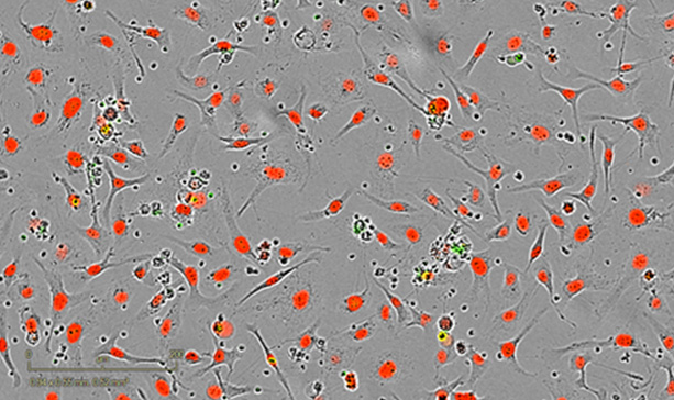
Treated – 24h post-coculture Treated – 24h post-coculture
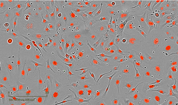
Control – 24h post-coculture Control – 24h post-coculture

SKOV3 ovarian tumor cells co-cultured with stimulated NK cells for 24 hours. Cetuximab exposure triggers ADCC, driving apoptotic death of antibody-coated tumor cells.

Imane Nafia, PhD, Chief Scientific Officer at Explicyte

The ADCC assay tells us whether an antibody can genuinely recruit NK cells to kill its target — not just bind it. By imaging tumor cell death in real time alongside IFNγ release, we separate true antibody-dependent killing from background NK activity, on the exact tumor model that matters for your program.

Imane Nafia, PhD

Chief Scientific Officer, Explicyte

Assay principle

How the ADCC assay works
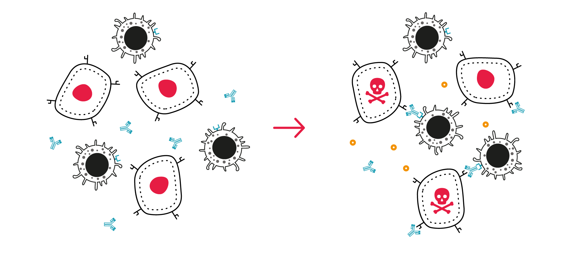
Diagram of the ADCC assay showing an antibody bridging an NK cell to a tumor cell, triggering tumor cell killing

1

Challenge tumor cells with candidate antibodies

Human tumor cell lines are exposed to test antibodies targeting a specific tumor antigen, then co-cultured with primary NK cells.

2

Track the tumor and immune response in parallel

Real-time live-cell imaging monitors tumor cell count and death using a nuclear probe and a caspase 3/7 apoptosis reagent. IFNγ release is quantified in parallel as a surrogate marker of NK cell activation.

3

Segment and analyze automatically

Automated image segmentation extracts kinetic apoptosis and proliferation curves across the full time course.

illustrative ADCC data

NK cell-mediated killing: example results

Target and effector cell characterization

Flow cytometry confirms CD16 expression on primary NK cells isolated from PBMCs (A), and HER2/EGFR expression levels on ovarian SKOV3 (B) and lung A549 (C) tumor cell lines used in the assay.
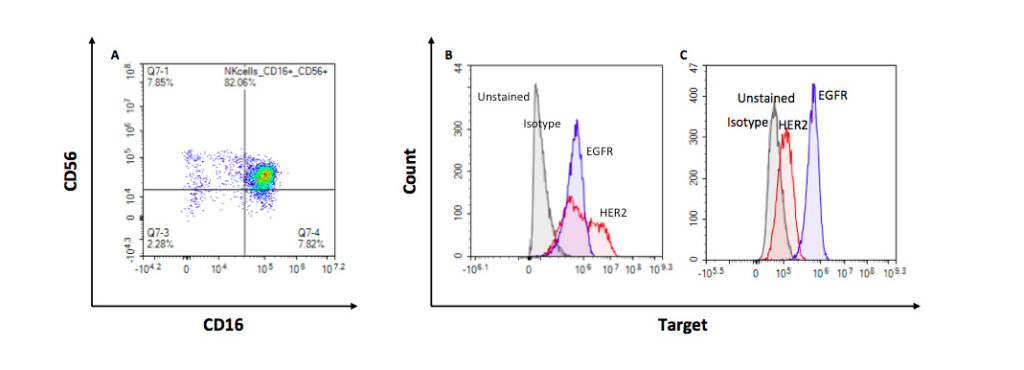
Flow cytometry histograms showing CD16 on NK cells and HER2/EGFR expression on SKOV3 and A549 tumor cells

IFNγ release confirms antibody-specific ADCC

SKOV3 (HER2+/EGFR+) and A549 (EGFR+/HER2−) tumor cells were treated with trastuzumab or cetuximab, then co-cultured with activated NK cells at increasing E:T ratios. IFNγ release, measured in supernatants 24 hours later, rose with NK ratio in all conditions — but cetuximab boosted it further on both cell lines, while trastuzumab did so only on HER2+ SKOV3. The assay correctly discriminates target-specific ADCC from basal NK activity.
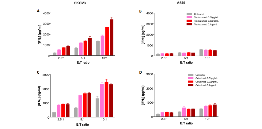
Bar chart of IFNγ release by activated NK cells treated with trastuzumab or cetuximab across E:T ratios

Avelumab drives NK-mediated killing of triple-negative breast cancer cells

MDA-MB-231 (TNBC) cells were treated with atezolizumab or avelumab, then co-cultured with activated NK cells at increasing E:T ratios. IFNγ release rose with NK ratio in all conditions, but only avelumab drove a further increase. Live-cell imaging confirmed the effect functionally, with avelumab-treated cells showing a corresponding rise in tumor cell apoptosis following NK addition. The assay distinguishes antibody-specific ADCC from basal NK activity, with cytokine and functional readouts in agreement.
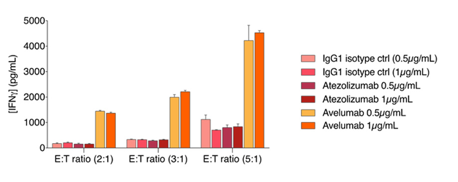
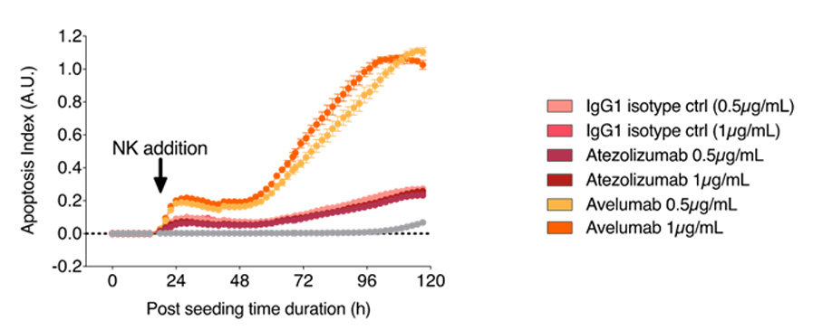
NK-mediated killing of MDA-MB-231 triple-negative breast cancer cells treated with avelumab ive-cell imaging of tumor cell apoptosis after avelumab treatment and NK cell addition

Making a difference as a preclinical CRO in oncology

Why work with Explicyte

Proven experience

>10 years in preclinical immuno-oncology, deep expertise in NK- and antibody-mediated killing, over 100 in vitro campaigns delivered.

More about us →

Built around your program

A PhD-level study director designs your study around your antibody, target antigen, and tumor model (over 100 tumor cell lines available).

Learn about our workflow →

Beyond functional readouts

A multiomic platform to resolve mechanism of action, alongside the core killing data — flow cytometry, proteomics, scRNAseq, Xenium-STAMP.

View our platform →

Reports you can act on

Progress reports let you adjust the plan as the study runs; final reports arrive with ready-to-publish data that holds up in due diligence.

See our publication record →

The Explicyte team at their Bordeaux laboratory

Paul Marteau, PharmD (study director), Imane Nafia, PhD (CSO), Loïc Cerf, MSc (COO), Alban Bessede, PhD (founder, CEO), Jean-Philippe Guégan, PhD (CTO)

Contact our team

Discuss your ADCC assay

Tell us about your antibody, its target antigen, and the tumor models you're considering — we'll scope the right assay design, readouts, and reporting, and send a fit-for-purpose proposal.

Answers about ADCC assay services

Frequently asked questions

What is an ADCC assay used for?

It measures whether a candidate antibody can direct NK cells to kill antibody-coated tumor cells — a core mechanism behind several approved monoclonal antibody therapies, including trastuzumab and cetuximab.

Two ways in parallel: real-time live-cell imaging tracks tumor cell death and proliferation directly, while IFNγ release in the culture supernatant serves as a functional readout of NK cell activation.

Yes. The assay is built around your candidate antibody and the tumor antigen it targets, using a tumor cell line expressing that antigen at appropriate levels.

Cell lines are selected to match your target antigen — SKOV3 and A549 are common examples for HER2 and EGFR. NK cells are primary cells isolated from human PBMCs, not an immortalized line.

Yes — cytokine release profiling, NK cytotoxic factor assessment (perforin/granzymes), proteomics, and transcriptomics are all available on request, alongside the core apoptosis and IFNγ readouts.

Each study returns kinetic apoptosis and proliferation curves for treated versus control conditions across the full time course, IFNγ quantification as a functional readout of NK cell activation, and dose-response and E:T ratio analysis across your candidate antibodies. You also receive a full study report with raw data, figures, and a statistical summary.

Explicyte Oncology CRO logo

Capabilities

Modalities