Immune Function & Modulation Assays

Mixed Lymphocyte Reaction (MLR) Assay

Does your compound modulate the dendritic-cell/T-cell immune synapse — amplifying an anti-tumor T-cell response, or relieving immunosuppression?

Our allogeneic MLR assay reads IFN-γ release as a surrogate of CD4+ T-cell activation, benchmarked against checkpoint inhibitors and adenosine-axis reference compounds.

MLR assay format and readouts
Format
96-well plate
Model
Allogeneic co-culture — primary CD4+ T cells (responder) with monocyte-derived dendritic cells (stimulator)
Readouts
IFN-γ release by HTRF as a surrogate of T-cell activation
Standards
Checkpoint inhibitors (anti-PD-1 nivolumab, anti-PD-L1 atezolizumab) and adenosine-receptor ligands (agonist to suppress, antagonist to relieve)
On request
Dendritic-cell immunophenotyping by flow cytometry, IL-8/IL-12/IL-10 for DC function, proteomics, and transcriptomics
MLR assays immuno-oncology CRO services

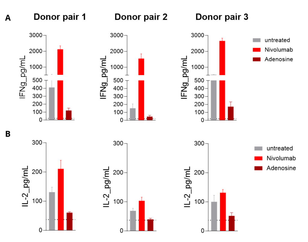
MLR in allogeneic human mDC/CD4+ T-cell co-cultures from independent donor pairs is enhanced by PD-1 inhibition and limited by adenosine. Peripheral-blood monocyte-derived mature dendritic cells are co-cultured with allogeneic CD4+ T cells; IFN-γ (A) and IL-2 (B) released into the supernatants are quantified by HTRF. The T-cell–stimulating capacity of mature DCs rises after PD-1 blockade with nivolumab (a higher MLR response) and is sharply reduced under adenosine treatment.

Imane Nafia, PhD, Chief Scientific Officer at Explicyte
The MLR shows whether a compound actually shifts the dendritic-cell/T-cell dialogue, not just whether it binds. Reading IFN-γ across independent donor pairs, against both checkpoint and adenosine-axis controls, tells us where in the synapse a candidate acts — and whether that effect holds beyond a single donor.

Imane Nafia, PhD

Chief Scientific Officer, Explicyte

Assay principle

How the MLR assay works
MLR assay CRO services cancer immunotherapy in vitro cell-based assays

1

Establish the allogeneic co-culture

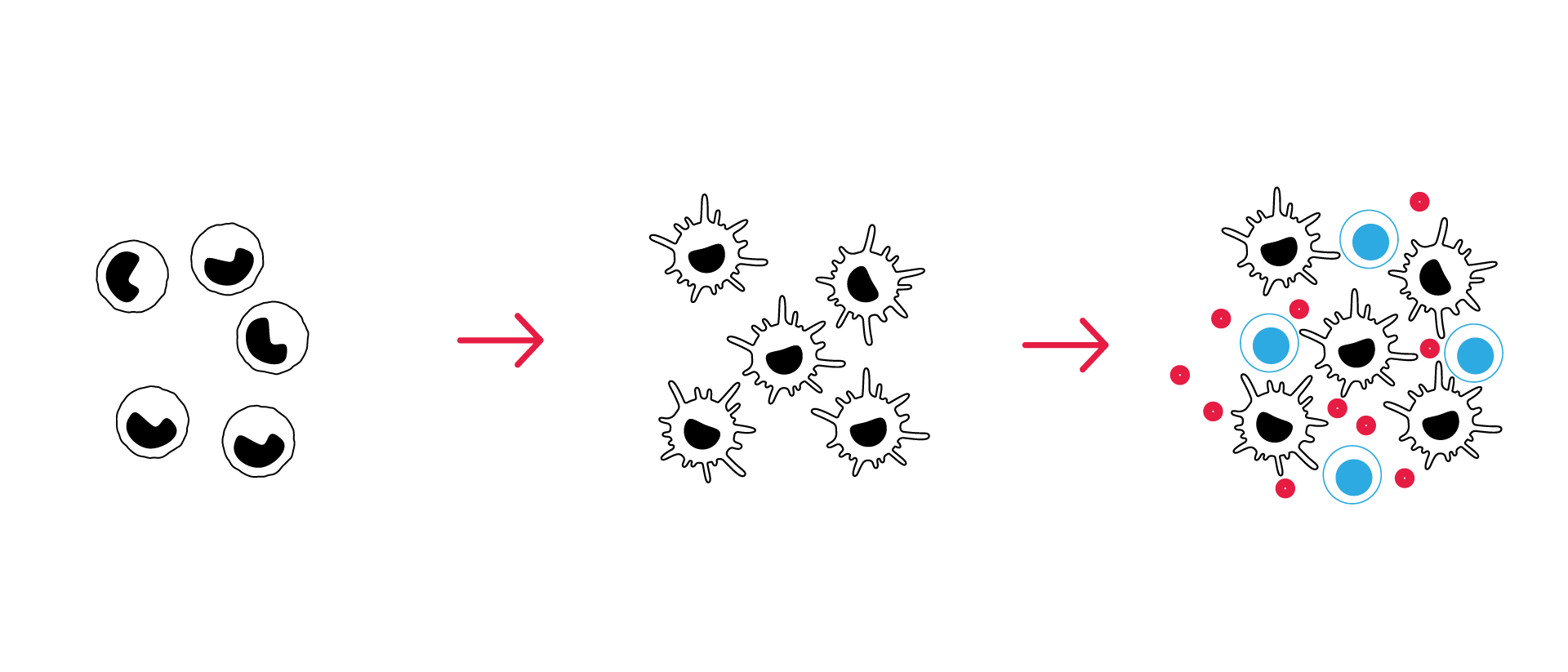
Monocyte-derived dendritic cells from one donor (stimulator) are co-cultured with CD4+ T cells from a second, unrelated donor (responder) in 96-well plates, driving an allogeneic T-cell response.

2

Introduce the test compound

Your compound is added during dendritic-cell differentiation and/or maturation, or at the co-culture stage — letting you probe where in the dendritic-cell/T-cell synapse it acts.

3

Quantify the T-cell response

Cytokines released during DC maturation and co-culture — IFN-γ, IL-2, IL-8, IL-12, IL-10 — are measured by HTRF or LEGENDplex™, with IFN-γ read as the primary surrogate of T-cell activation.

4

Resolve the mechanism of action

Surface-marker phenotyping and signaling-pathway analysis pin down how the compound shifts dendritic-cell function and the resulting T-cell response.

Illustrative MLR data

MLR assay for cancer immunotherapies: example results

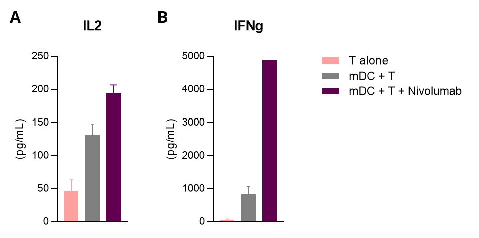
PD-1 blockade enhances the allogeneic MLR response

Monocyte-derived mature dendritic cells co-cultured with allogeneic CD4+ T cells; IL-2 (A) and IFN-γ (B) in the supernatants quantified by HTRF. Mature DCs drive a measurable MLR response that increases further after PD-1 blockade with nivolumab.
MLR assay data — PD-1 blockade with nivolumab increases IL-2 and IFN-γ release in allogeneic mDC/CD4+ T-cell co-culture

Adenosine impairs dendritic-cell function, relieved by receptor antagonism

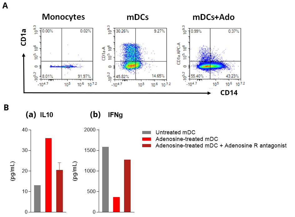
Monocyte-derived DCs were challenged with adenosine, with or without an adenosine-receptor antagonist. Flow cytometry profiled DC differentiation and maturation (A); DC and co-culture supernatants were assayed by HTRF for IL-10 and IFN-γ (B).

Adenosine skewed DCs toward a CD1a-low CD14+ phenotype and suppressed the MLR — more IL-10 from DCs, less IFN-γ from T cells — an effect reversed by the antagonist, confirming it is adenosine-receptor–mediated.
MLR assay data — adenosine skews dendritic-cell phenotype and suppresses the MLR, reversed by an adenosine-receptor antagonist

Making a difference as a preclinical CRO in oncology

Why work with Explicyte

Deep immuno-oncology experience

>100 in vitro campaigns over the past ten years, and 30+ peer-reviewed publications in immuno-oncology journals.

See our latest papers →

A dedicated PhD study director

One study director, PhD-level, owns your program from experimental design through the final report discussion.

Learn about our workflow →

Independent donor pairs

Characterized donors and controlled differentiation, run across unrelated responder–stimulator pairs so an effect holds beyond one donor.

See our publication record →

Mechanism, not just a readout

Immunophenotyping, cytokine multiplexing, proteomics, and transcriptomics locate where your compound acts — on DC function, the T-cell response, or both.

View our platform →

The Explicyte team at their Bordeaux laboratory

Paul Marteau, PharmD (study director), Imane Nafia, PhD (CSO), Loïc Cerf, MSc (COO), Alban Bessède, PhD (founder, CEO), Jean-Philippe Guégan, PhD (CTO)

Contact our team

Discuss your MLR assay

Whether you're testing an immune checkpoint inhibitor, an agonist antibody, or a small molecule acting on an immunosuppressive pathway, we'll build an MLR study around your mechanism and endpoints — from single-agent screening to combination potency profiling.

Answers about MLR assay

Frequently asked questions

What does a mixed lymphocyte reaction (MLR) assay measure?

It measures how a test compound modulates the dendritic-cell/T-cell immune synapse. In an allogeneic co-culture of CD4+ T cells (responder) and monocyte-derived dendritic cells (stimulator), IFN-γ release reports the strength of the resulting T-cell activation — rising when a compound amplifies the response and falling when it suppresses it.

Primary CD4+ T cells from one donor and monocyte-derived dendritic cells from a second, unrelated donor, co-cultured in 96-well plates. The unrelated donors create the allogeneic mismatch that drives the T-cell response.

IFN-γ by HTRF is the primary readout, as a surrogate of T-cell activation. Depending on the question, we also measure IL-2, IL-8, IL-12 and IL-10, phenotype the dendritic cells by flow cytometry, and add proteomic or transcriptomic profiling to resolve mechanism.

Checkpoint inhibitors — anti-PD-1 (nivolumab) and anti-PD-L1 (atezolizumab) — as amplifying controls, and adenosine-receptor ligands as the immunosuppressive axis: an agonist to suppress the response, an antagonist to relieve it.

Both biologics and small molecules, as single agents or in combination — including immune checkpoint inhibitors, agonist antibodies, and compounds acting on immunosuppressive pathways such as the adenosine axis.

Yes. Compounds can be introduced during dendritic-cell differentiation, during maturation, or at co-culture, and DC phenotype can be read before co-culture. That separates effects on dendritic-cell function from effects on the T-cell response itself.

The assay is run across independent donor pairs, so a compound’s effect is observed beyond a single responder–stimulator combination rather than in one genetic background.

Screening, potency profiling, and functional characterization of candidate compounds — from an initial single-agent read through combination studies with immune checkpoint inhibitors.

Explicyte Oncology CRO logo

Capabilities

Modalities