Tumor killing assays

T Cell–Mediated Tumor Killing Assays

Does your candidate increase T-cell-mediated tumor killing, by activating T cells, sensitizing tumor cells or redirecting cytotoxicity toward a defined target?

We co-culture human tumor cells with primary immune cells and monitor tumor-cell growth and apoptosis in real time, alongside functional immune readouts. The assay can be tailored to checkpoint inhibitors, immune agonists, tumor-sensitizing compounds and T-cell engagers.

A flexible functional platform for a broad range of T-cell-directed therapies and mechanisms.

T cell killing assay format and readouts
Format
96- or 384-well plates; dose-response
Model
Human tumor cell lines (>100 cell lines) co-cultured with primary immune cells (PBMCs or purified T / CD8 cells)
Core readouts
Tumor-cell count (nuclear fluorescent probe) + apoptosis (caspase-3/7 reagent) by live-cell imaging (Incucyte), plus a soluble marker of immune activation by HTRF — IFN-γ in most models, with TNF-α, IL-6, or other cytokines selected to fit the biology.
Standards
Benchmarked with clinically-validated immune checkpoint inhibitors, with study-specific controls selected according to modality and mechanism of action.
On request
Flow-cytometry immunophenotyping, cytokine / secretome (HTRF, LEGENDplex), proteomics, transcriptomics
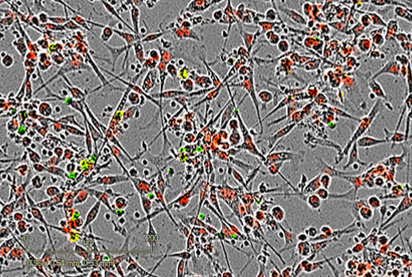
72h post-treatment 72h post-treatment
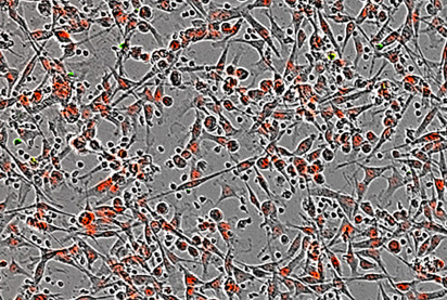
Untreated Untreated

Inhibition of a specific tumor target sensitizes breast MDA-MB-231 tumor cells to immune cell-mediated killing. Breast cancer MDA-MB-231 cells were seeded, then treated or not with a tumor target inhibitor. They were then co-cultured with activated PBMCs. Tumor cell proliferation (count) & apoptosis were monitored and analyzed as surrogate measures of immune cell-mediated tumor cell death. Image fields are representative of tumor proliferation / apoptosis at 72h post-coculture.

Imane Nafia, PhD, Chief Scientific Officer at Explicyte

Real-time imaging shows whether tumor control results from reduced growth, increased apoptosis or both. By combining these kinetic readouts with immune activation and cytotoxic-effector profiling, we can link the functional outcome to the mechanism driving it.

Imane Nafia, PhD

Chief Scientific Officer, Explicyte

Assay principle

How the T cell killing assay works
T cell activation immune checkpoint inhibition killing assays CRO in vitro services

1

Set up the co-culture

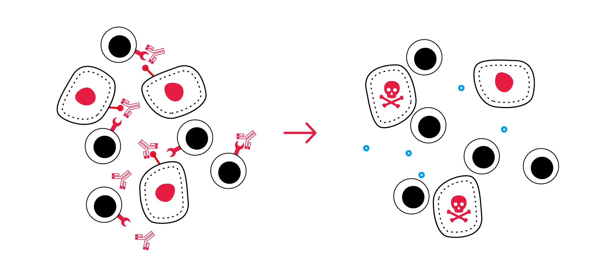
Co-culture human tumor cells with primary PBMCs, T cells, or CD8 T cells at an optimized effector-to-target ratio. The candidate is applied to tumor cells, immune cells, or the complete co-culture according to its mechanism.

2

Monitor tumor killing and immune activation

Track tumor-cell count and apoptosis over 3–5 days by live-cell imaging, while measuring selected soluble markers of immune activation .

3

Resolve the mechanism

Add flow cytometry, cytokine profiling, proteomics, or transcriptomics to characterize immune activation, cytotoxic effector function, and mechanism of action.

Discuss your program

IMMUNE-MEDIATED TUMOR KILLING: ILLUSTRATIVE DATA

T-cell killing under checkpoint blockade

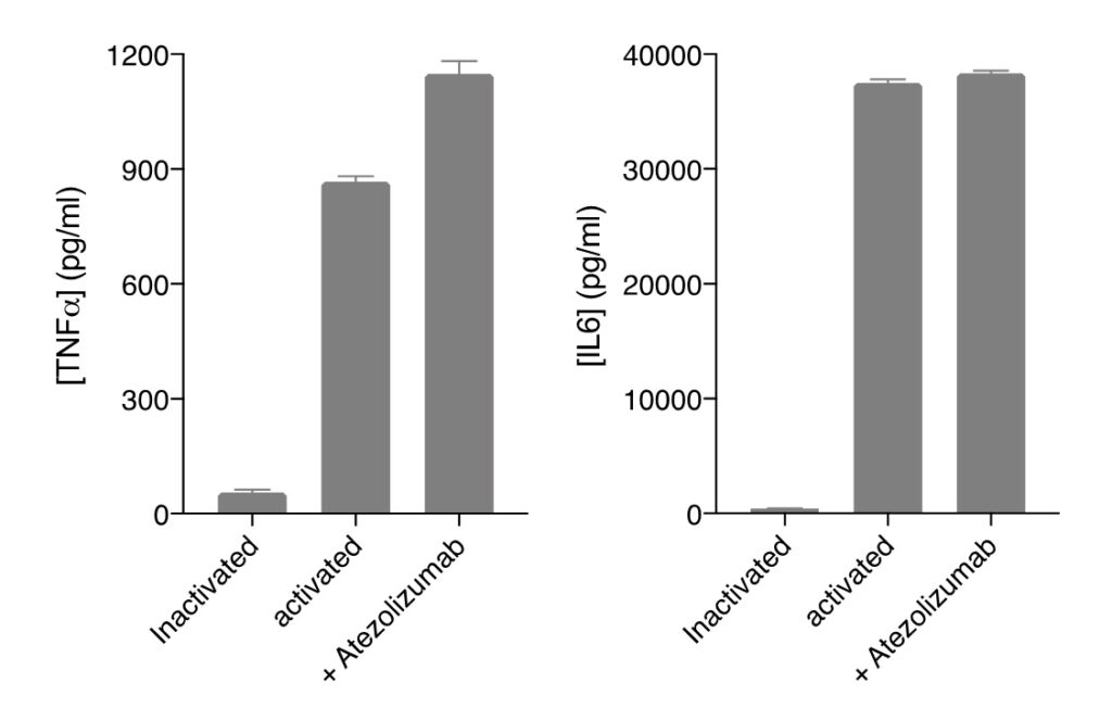
Anti-PD-L1 mAb (Atezo) promotes immune cell-mediated killing of breast MDA-MB-231 tumor cells.

MDA-MB-231 tumor cells were cultured either alone (ctrl) or with PBMCs in the presence and absence of PD-L1 inhibitor (Atezo). Supernatants were collected at 72h post-co-culture and analyzed by HTRF for TNF⍺ and IL6 release as surrogates of immune cell response (A). Tumor cell count was also kinetically monitored, normalized, and analyzed as surrogate of tumor cell proliferation and growth (B). While tumor cell exposure to activated immune cells is sufficient to induce an immune cell response against MDA-MB-231 tumor cells as illustrated by the increase of TNF⍺ levels (A) and decrease of the tumor cell number (B), Atezo is shown to further optimize the immune cell-mediated killing activity.

A. Atezo promotes immune cell response
Atezo promotes immune cell response
B. Atezo promotes anti-tumor immune cell-mediated activity
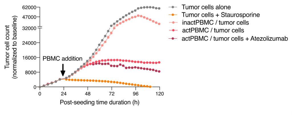
Atezo promotes anti-tumor immune cell-mediated activity

Activated immune cell-mediated killing of A549 lung cancer cells is enhanced by PDL1 blockade.

Lung cancer A549 cells were seeded and then co-cultured – at an appropriate E:T ratio – with activated hPBMCs, in the presence and absence of Atezo. (A) Tumor cell apoptosis was monitored over ~5 days by mean of a caspase 3/7 fluorescent probe and a NucRED probe expression, respectively, as a surrogate measure of immune cell killing activity towards tumor cells, and quantified in kinetics (left) or as AUC (right). (B) Tumor cell count was monitored as a surrogate of tumor cell proliferation, and quantified in kinetics (left) or as AUC (right).
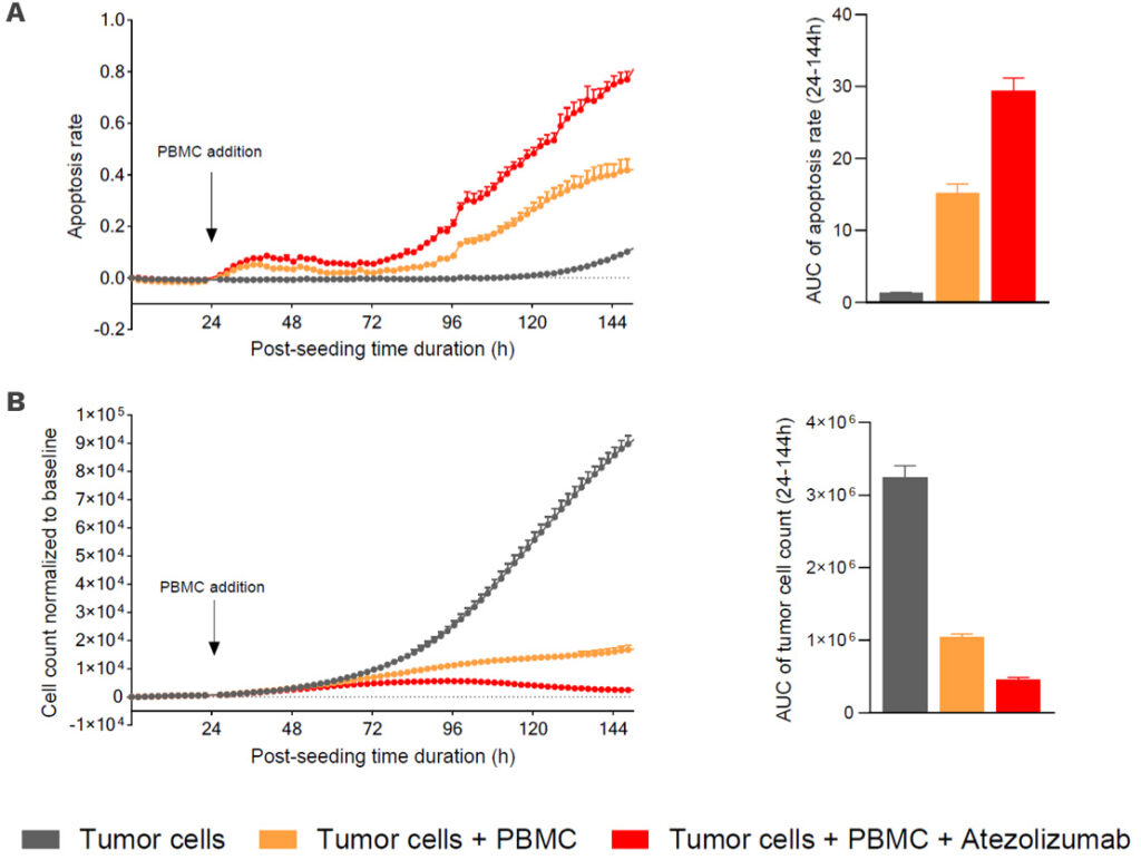
immune checkpoint blockade PBMC tumor killing

CD8 T cell-mediated killing of A375 melanoma cells is promoted by increasing anti-CD3 concentration

Melanoma A375 tumor cells were seeded and 48h later were co-cultured with activated CD8 cells, in the presence and absence of increasing concentrations of anti-CD3 antibody. (A) Tumor cell apoptosis was monitored and quantified over a period of ~3 days, by mean of a caspase 3/7 fluorescent probe and a NucRED probe expression, respectively, as surrogate measures of immune cell killing activity towards tumor cells. (B) IFNg released levels in the co-culture supernatants were quantified by mean of a specific HTRF detection kit. Data are expressed as means ± SEM.
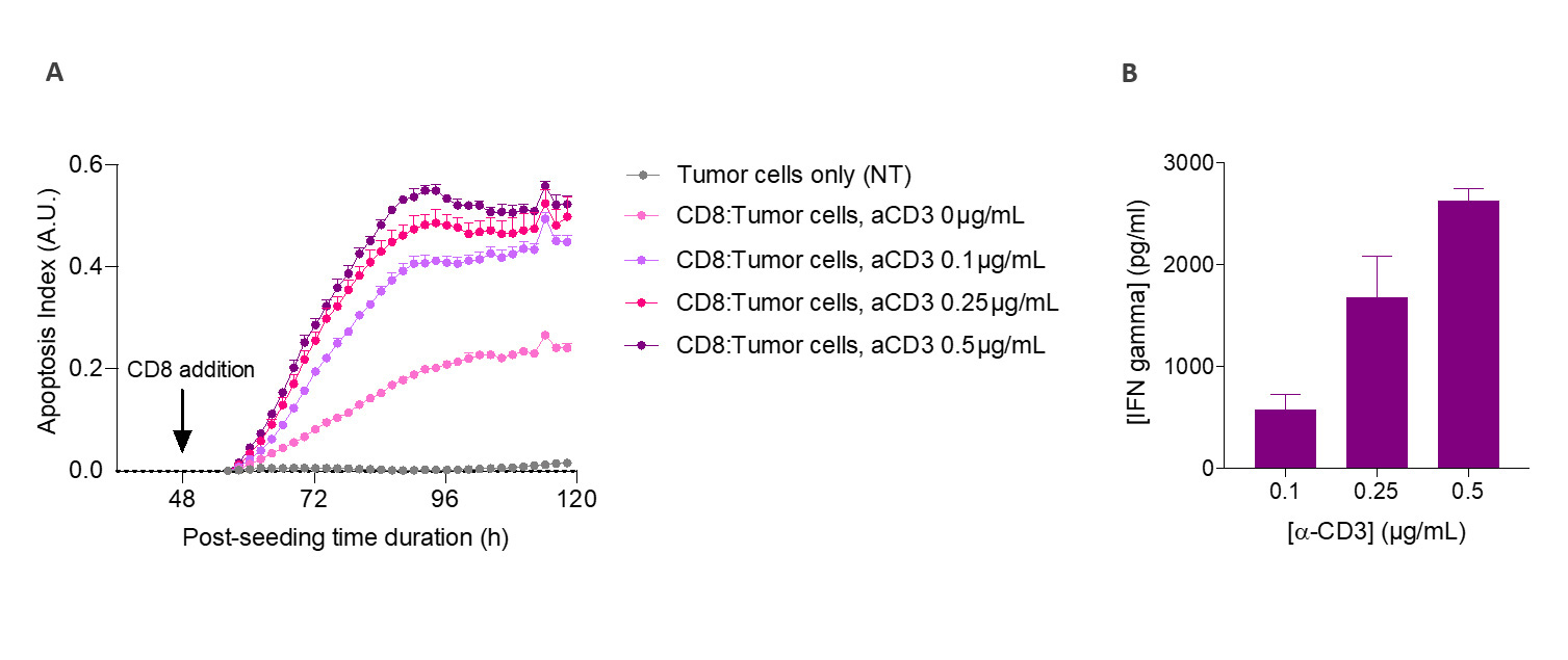
T-cell killing assay CRO in vitro screening CD8 T A375 melanoma cells
Discuss your program

T-cell engager case study

T-cell engagers redirect killing onto the target
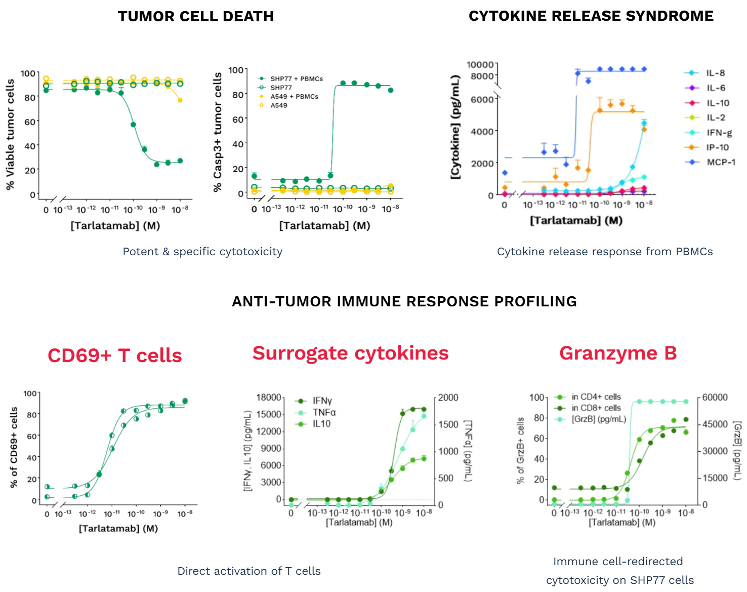
A DLL3-targeted T-cell engager kills on-target and spares off-target

After selecting cell lines that express DLL3 (SHP77) or not (A549, control), we assessed tarlatamab in dose-response, co-cultured with primary human T cells.

Potency & specificity: Tarlatamab induced tumor-cell death — fewer viable cells, more caspase-3/7-positive cells — on DLL3-positive SHP77 in the presence of PBMCs only. DLL3-negative A549 cells were spared.

Anti-tumor immune response: T-cell activation (CD69+), surrogate cytokines (IFN-γ, TNF-α), and cytotoxic effector molecules (granzyme B) rose dose-dependently, redirecting immune killing onto SHP77 cells.

Cytokine release: A cytokine release response from PBMCs — IL-8, IP-10, MCP-1, IFN-γ and others — was profiled alongside the kill.

In vitro profiling of DLL3-targeted BiTE Tarlatamab
Discuss your program

Making a difference as a preclinical CRO in oncology

Why work with Explicyte

Proven experience

>10 years in preclinical immuno-oncology, deep expertise in T-cell killing assays, over 100 in vitro campaigns delivered.

More about us →

Built around your program

A PhD-level study director designs the study around your target, tumor model, and modality, from experimental plan to final-report discussion.

Learn about our workflow →

Beyond the kill

Immunophenotyping, cytokine and secretome analysis, proteomics, and single-cell / spatial transcriptomics resolve the mechanism behind the functional result.

View our platform →

Reports you can act on

Progress reports let you adjust the plan as the study runs; final reports arrive with ready-to-publish data that holds up in due diligence.

See our publication record →

The Explicyte team at their Bordeaux laboratory

Paul Marteau, PharmD (study director), Imane Nafia, PhD (CSO), Loïc Cerf, MSc (COO), Alban Bessede, PhD (founder, CEO), Jean-Philippe Guégan, PhD (CTO)

Contact our team

Discuss your tumor killing study

Tell us your target, the modality you're testing (checkpoint, sensitizer, or T-cell engager), and the tumor indication — we'll scope the right model, effector cells, readouts, and controls, and send a fit-for-purpose proposal.

Answers about T-cell killing assays

Frequently asked questions

What is a T cell–mediated tumor killing assay?

An in vitro co-culture assay that measures how primary human immune cells affect tumor-cell growth and survival over time. Tumor-cell count and apoptosis are monitored by live-cell imaging, alongside selected immune-response readouts. Purified T-cell or CD8 T-cell formats can be used when a more focused assessment of T-cell-mediated activity is required.

Any modality whose mechanism runs through T-cell-mediated killing — immune checkpoint inhibitors, T-cell agonists and co-stimulators, tumor-sensitizing agents, and bispecific / multispecific T-cell engagers — alone or in combination, including with standard-of-care checkpoint antibodies. If your candidate is meant to make T cells kill tumor cells, the assay can be designed around it.

Primary human PBMCs, purified T cells, or CD8 T cells, co-cultured with human tumor cell lines from a library of 100+ (breast, lung, melanoma, small-cell lung cancer, and others) at an optimized effector-to-target ratio.

Core readouts include tumor-cell count and caspase-3/7-dependent apoptosis monitored kinetically over approximately 3–5 days. Tumor-cell count reflects net tumor growth, while caspase-3/7 provides a complementary measure of apoptosis. Soluble markers such as IFN-γ, TNF-α or IL-6 are selected according to the immune cell format and study biology. Optional readouts include flow-cytometry immunophenotyping, cytokine and cytotoxic-effector profiling, proteomics and transcriptomics.

The dose-response kill is run on a target-positive tumor line alongside a target-negative control line (both confirmed by flow cytometry). A specific engager kills the target-positive line only in the presence of immune cells, and spares the target-negative line — as shown in our tarlatamab (DLL3) case study.

Benchmark standards include anti-PD-1 (nivolumab, pembrolizumab) and anti-PD-L1 (atezolizumab, avelumab), with anti-CD3 as an activation reference. Sensitization studies compare treated versus untreated tumor cells; engager studies use a target-negative control line.

We work from a library of 100+ human tumor cell lines spanning solid and hematological indications, so most targets and indications can be covered — and we can source or bring in a specific line on request. Lines used in the examples on this page include MDA-MB-231 (breast), A549 & SHP77 (lung), and A375 (melanoma).

96- or 384-well plates in dose-response format, with kinetic imaging reads over roughly 3–5 days. Timeline depends on the number of conditions, cell lines, and add-on readouts — a study director scopes it with you.

Explicyte Oncology CRO logo

Capabilities

Modalities