Tumor killing assays

Tumor antigen-specific T cell killing assays

Does your vaccine or antigenic candidate generate a functional tumor-specific CD8 T-cell response? Or can your compound enhance dendritic-cell antigen presentation and the priming of tumor-reactive T cells?

This assay links antigen presentation to CD8 T-cell activation and antigen-specific tumor killing, enabling the evaluation of both antigenic candidates and immune-modulating compounds within a defined antigen–tumor system.

Tumor antigen-specific T cell killing: format and readouts
Format
Miniaturized DC cultures, DC–CD8 co-cultures, and CD8–tumor co-cultures.
Model
Human DC-primed CD8 T cells co-cultured with antigen-matched target tumor cells.
Readouts
Tumor-cell apoptosis and count by live-cell imaging, plus IFN-γ release as the T-cell activation surrogate — IFN-γ in most models; other cytokines as the biology fits.
Reference
A qualified Melan-A / SK-MEL-5 system is available as a positive reference model for antigen-specific CD8 T-cell priming and tumor-cell killing. For sponsor-defined antigens or vaccine candidates, study-specific controls are selected according to the antigenic format, HLA restriction and tumor model.
On request
Flow-cytometry immunophenotyping (DC differentiation and maturation markers included), cytokine release, proteomics, transcriptomics.
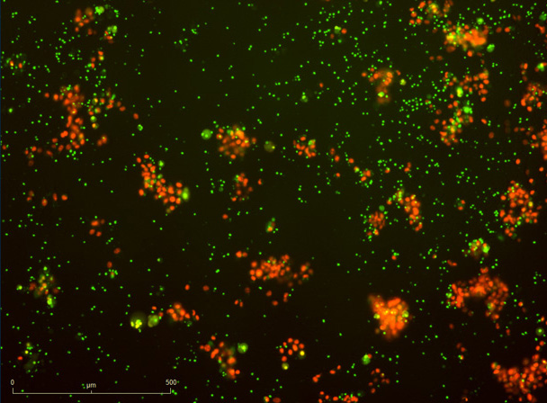
Killing after antigen-priming Killing after antigen-priming
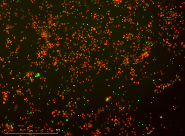
Killing after control priming Killing after control priming

DC-mediated priming of CD8 T cells drives antigen-specific killing of SK-MEL-5 (Melan-A⁺) melanoma cells. SK-MEL-5 targets were co-cultured with CD8 T cells primed by either untreated or Melan-A-treated dendritic cells. Tumor-cell count and apoptosis were tracked over ~72 h as surrogate measures of immune-cell-mediated killing (representative fields above).

Imane Nafia, PhD, Chief Scientific Officer at Explicyte

Antigen-specific killing only means something if you can see both halves — the priming that antigen presentation drives, and the tumor-cell death that follows. We read them in parallel, so the mechanism and the outcome come from the same experiment.

Imane Nafia, PhD

Chief Scientific Officer, Explicyte

Assay principle

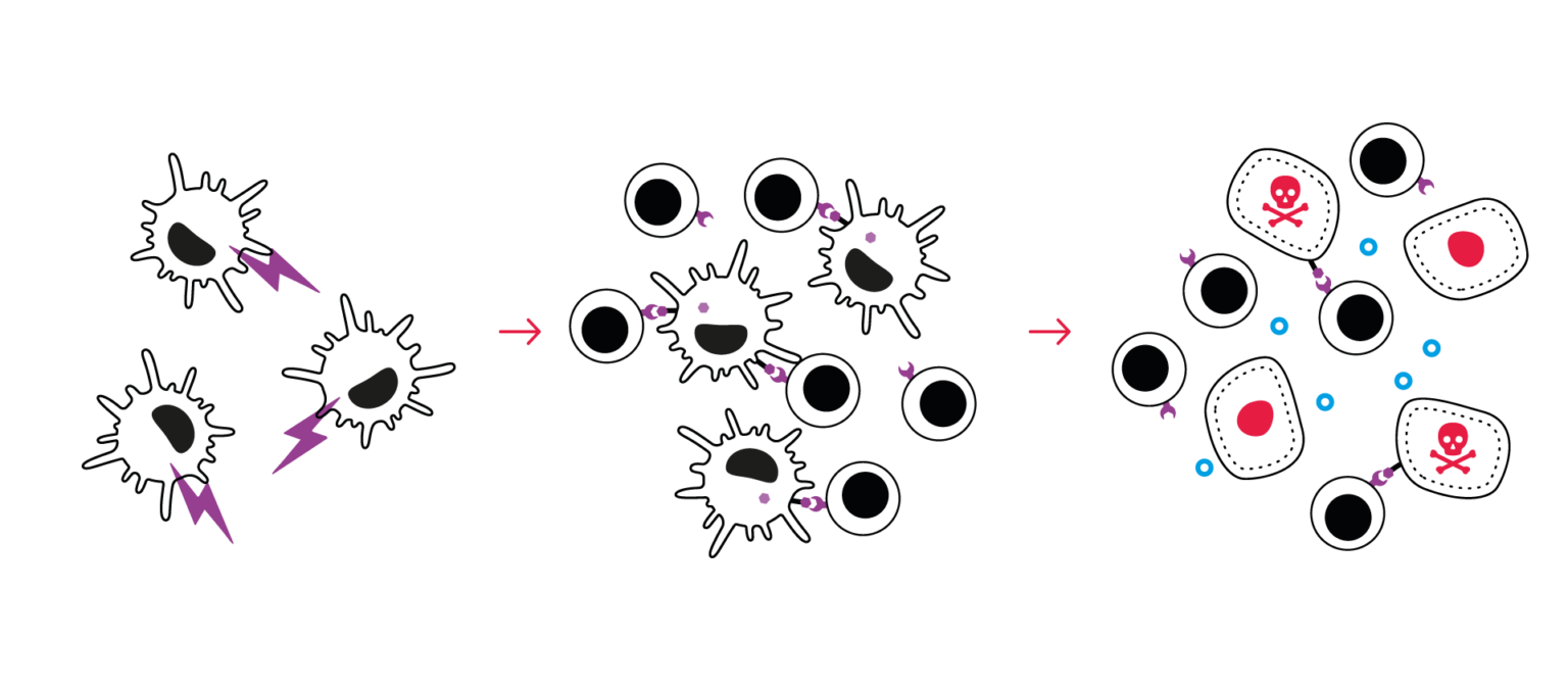
How the tumor antigen-specific T cell killing assay works
Tumor antigen specific T cell killing assay CRO immuno-oncology

1

Antigen loading and dendritic-cell modulation

Dendritic cells are exposed to the antigenic candidate, such as a peptide, vaccine construct or tumor-derived material. Alternatively, a defined antigen such as Melan-A can be used to evaluate compounds that enhance dendritic-cell maturation, antigen processing or presentation.

2

CD8 T-cell priming

Antigen-loaded dendritic cells are co-cultured with autologous CD8 T cells to generate a tumor-reactive T-cell response. Test compounds can also be added at this stage to assess their ability to improve dendritic-cell–T-cell interactions and CD8 T-cell priming.

3

Antigen-specific tumor-cell killing

Primed CD8 T cells are challenged with antigen-positive tumor cells. Tumor-cell apoptosis and growth are monitored by live-cell imaging, while cytokine release and optional immune profilingcharacterize the associated T-cell response.
Discuss your program

Antigen-specific killing case study

Enhancing DC antigen presentation drives the kill
Enhancing DC antigen presentation drives antigen-specific killing of SK-MEL-5

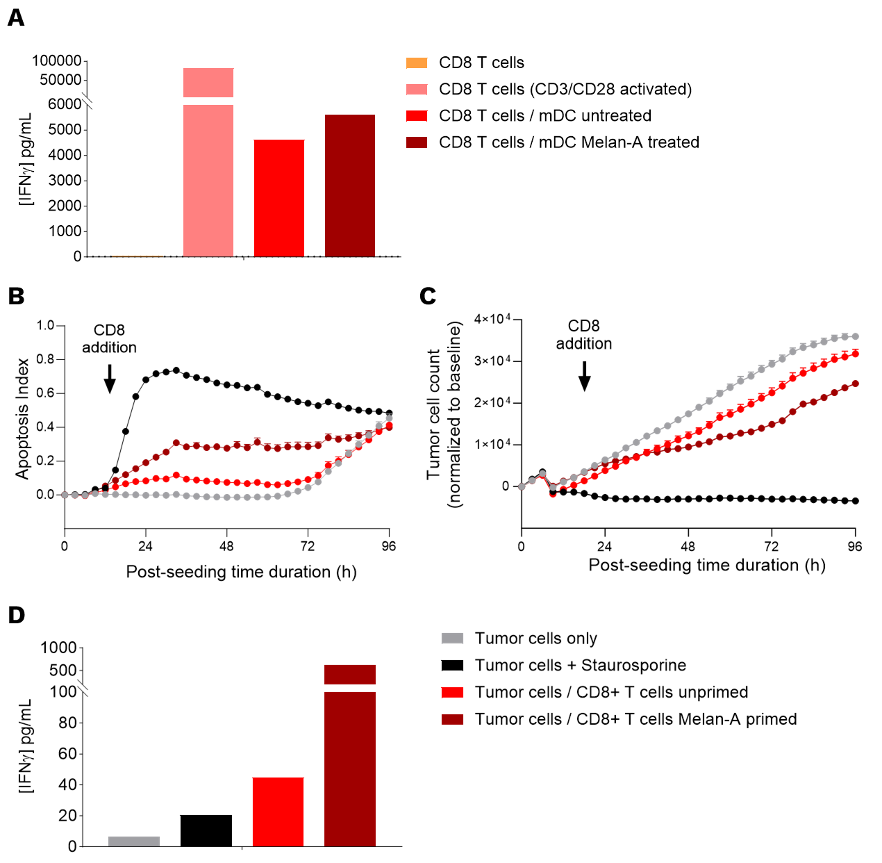
A dendritic-cell–primed model: treating DCs with the Melan-A tumor antigen sharpens CD8 priming and produces antigen-specific killing of Melan-A⁺ melanoma cells.

(A) Priming: Treating DCs with the Melan-A peptide during maturation improved CD8 T-cell priming, seen as higher IFN-γ in CD8 / mature-DC co-culture supernatants versus untreated DCs

(B, C) Antigen-specific killing: CD8 T cells primed by Melan-A-treated DCs killed SK-MEL-5 (Melan-A⁺) targets over ~4 days — higher apoptosis rate (B) and lower tumor-cell count (C) than CD8 primed by untreated DCs.

(D) Immune response: IFN-γ in the SK-MEL-5 / CD8 co-culture supernatants rose in step with the kill when CD8 were primed by antigen-treated DCs.

enhancement of DC antigen presentation T-cell killing assay
Discuss your program

Making a difference as a preclinical CRO in oncology

Why work with Explicyte

A decade of in-vitro immuno-oncology

>100+ in-vitro campaigns, 30+ peer-reviewed publications in immuno-oncology journals, across the mechanisms that antigen-specific killing depends on.

More about us →

Built around your program

One study director (PhD level) from experimental design through final-report discussion, shaping the readouts and treatment windows around your mechanism of action.

Learn about our workflow →

Mechanism, not just a number

Add immunophenotyping, cytokine release, proteomics, or transcriptomics to read the DC and T-cell components behind the kill.

View our platform →

Reports you can act on

Progress reports let you adjust the plan as the study runs; final reports arrive with ready-to-publish data that holds up in due diligence.

See our publication record →

The Explicyte team at their Bordeaux laboratory

Paul Marteau, PharmD (study director), Imane Nafia, PhD (CSO), Loïc Cerf, MSc (COO), Alban Bessede, PhD (founder, CEO), Jean-Philippe Guégan, PhD (CTO)

Contact our team

Discuss your tumor killing study

Tell us your target, the modality you're testing (checkpoint, sensitizer, or T-cell engager), and the tumor indication — we'll scope the right model, effector cells, readouts, and controls, and send a fit-for-purpose proposal.

Answers about antigen-specific killing

Frequently asked questions

What does this assay measure?

The assay measures whether dendritic-cell-mediated antigen presentation generates a functional CD8 T-cell response capable of killing antigen-matched tumor cells. It can evaluate either the immunogenic potential of an antigenic candidate or the activity of a compound that modulates dendritic-cell function or T-cell priming.

The T cell–mediated killing assay reads redirected or polyclonal killing. This assay is antigen-driven: the effector CD8 population is primed through antigen-presenting cells you can treat, and specificity is read as killing of antigen-positive targets versus antigen-negative or unexposed-DC controls. The two are complementary; many programs run both.

Antigenic candidates may include peptides, protein- or nucleic-acid-based vaccine constructs, tumor-derived materials and oncolytic viruses. The assay can also evaluate compounds that enhance dendritic cell maturation, antigen processing or presentation, or productive DC–CD8 T-cell interactions within a defined antigen–tumor system.

Core readouts are tumor-cell apoptosis and count by live-cell imaging (nuclear probe for count, caspase-3/7 reagent for death) and IFN-γ release as the T-cell activation surrogate — IFN-γ in most models, with TNF-α, IL-6, or other cytokines added as the biology fits. On request: flow-cytometry immunophenotyping (including DC differentiation and maturation markers), broader cytokine panels, proteomics, and transcriptomics.

The assay can be adapted to a wide range of tumor antigens and antigenic formats, including peptides, proteins, vaccine constructs, tumor-derived material and other sponsor-defined candidates. Tumor models are selected according to antigen expression, HLA compatibility and relevance to the intended indication. Autologous dendritic cells and CD8 T cells are then combined with the selected antigen-matched tumor target. A Melan-A / SK-MEL-5 system is available as a qualified reference model.

Specificity is assessed by comparing CD8 T cells primed with antigen-exposed versus control dendritic cells and, where available, antigen-positive versus antigen-negative tumor targets. Additional controls may include CD8-only, tumor-only, and immune-cell-free conditions. Immune-modulating reference compounds can be included when relevant to the mechanism being studied.

Starting from FFPE cohorts, our translational team can explore or confirm target-antigen expression across cancer indications using single-cell RNA-seq, Xenium single-cell spatial transcriptomics, GeoMx digital spatial profiling, and digital pathology. See target expression profiling.

A fit-for-purpose study design agreed up front, a dedicated PhD study director throughout, and a final report with the quantified killing and activation readouts, the requested mechanism add-ons, and a discussion of the results against your objectives.

Explicyte Oncology CRO logo

Capabilities

Modalities