Immune Function & Modulation Assays

M1 & M2 macrophage assays

Can your compound repolarize M2 macrophages to M1, or relieve M2-mediated immunosuppression?

Our M1/M2 assays read that out with IFN-γ as the surrogate of T cell activation.

Macrophage assays' format and readouts
Format
Miniaturized culture plates
Models
Monocyte-derived M1 or M2 macrophages, alone or co-cultured with autologous activated PBMCs or CD4+ T cells
Readouts
IFN-γ as a surrogate of T cell activation; macrophage cytokine release (IL-6, IL-10, IL-12)
Standards
p38 MAPK inhibitor, STAT3 inhibitor, PI3K inhibitor; nivolumab (anti-PD-1), atezolizumab (anti-PD-L1), ipilimumab (anti-CTLA-4)
On request
Extended macrophage and effector-cell cytokine profiling, flow-cytometry immunophenotyping, proteomics, transcriptomics
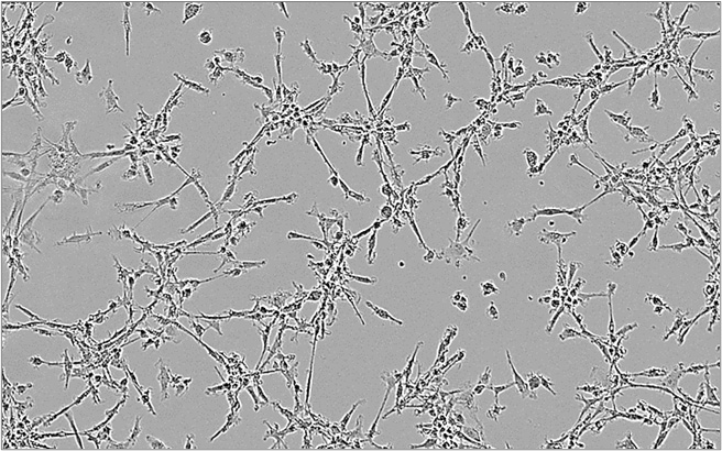
M2 macrophages M2 macrophages
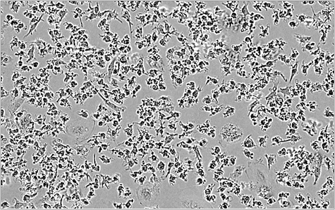
M1 macrophages M1 macrophages

Macrophages span functionally opposite states: M1 drive inflammation, while M2 resolve it and suppress T cells — through checkpoint engagement (e.g., PD-L1), anti-inflammatory cytokines (IL-10, TGF-β), and immunosuppressive metabolism, the same playbook as tumor-associated macrophages (TAMs). Our M2 suppression assay co-cultures monocyte-derived M2 macrophages with activated CD4+ T cells and reads IFN-γ to score how a compound modulates them.

Imane Nafia, PhD, Chief Scientific Officer at Explicyte

Getting a genuine, stable M2 phenotype is where most macrophage work falls down. We anchor polarization to CD86, CD163, and the IL-6/IL-10 balance, so when a compound shifts the state we know it’s the drug, not drift in the model.

Imane Nafia, PhD

Chief Scientific Officer, Explicyte

Assay principle

How our macrophage assays work
M2 Macrophage-Suppression Assay CRO immuno-oncology immunotherapy

1

Differentiate & polarize

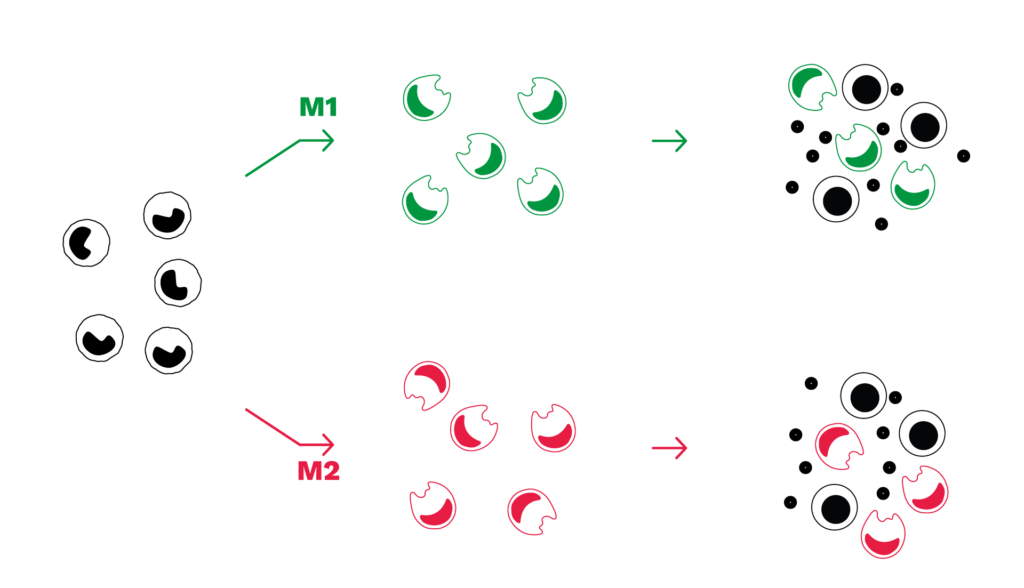
Monocytes are isolated from healthy-donor PBMCs and polarized in vitro into M1 or M2 macrophages.

2

Co-culture with T cells

M1 or M2 macrophages are co-cultured with autologous activated PBMCs or CD4+ T cells. Test compounds can be added during macrophage polarization, during the co-culture, or across both windows — as single agents or in combination.

3

Read out

IFN-γ is quantified as a surrogate of T cell activation, alongside macrophage cytokine release (IL-6, IL-10, IL-12).

4

Interpret

Effects are benchmarked against reference standards — pathway inhibitors and checkpoint antibodies — to place your compound’s repolarizing or suppression-relieving activity.

Illustrative macrophage data

Macrophage polarization and M2 suppression: example results

Mirrored M1/M2 phenotypes

M1 and M2 macrophages are functionally opposite subsets with mirrored phenotypes: M1 are CD86-high, CD163-negative, IL-6-high, IL-10-low; M2 are CD86-low, CD163-positive, IL-6-low, IL-10-high.
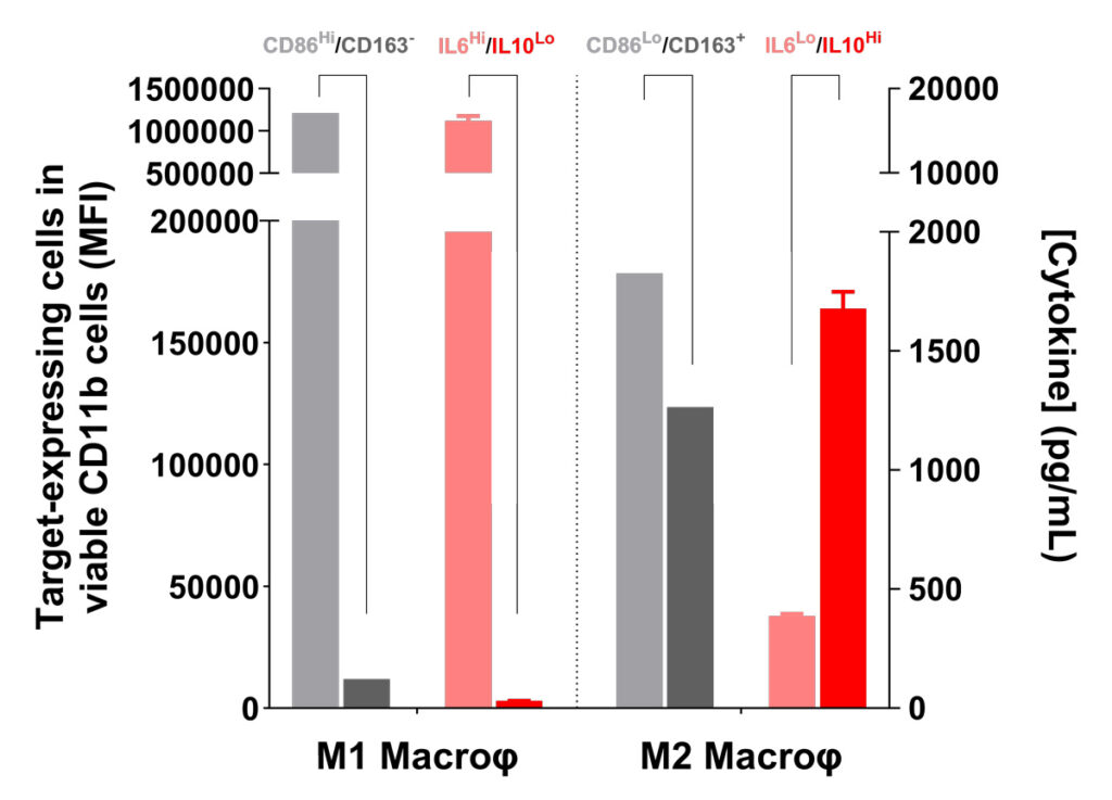
Flow cytometry marker expression and cytokine profile of M1 and M2 macrophages

Repolarizing M2 through signaling pathways

The M2 IL-6-low / IL-10-high profile drives T cell suppression via JAK/STAT and SMAD/p38 MAPK.

Adding a STAT3 or p38 MAPK inhibitor during polarization reverses that profile and relieves M2 suppression, seen as recovered IFN-γ in PBMC/M2 co-culture.
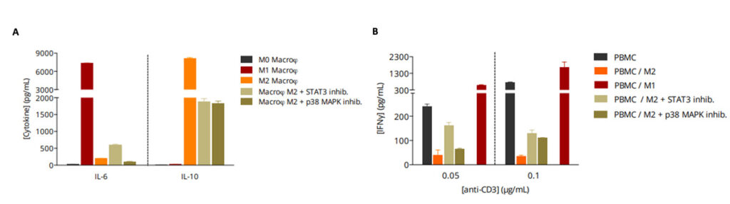
STAT3 and p38 MAPK inhibition repolarize M2 macrophages and restore IFN-γ

Relieving M2 suppression with checkpoint blockade

M2 suppression runs through PD-1/PD-L1 and CTLA-4/CD80–CD86. Nivolumab, atezolizumab, or ipilimumab restore T cell activation in PBMC/M2 co-cultures, and further promote the M1-driven stimulation of T cell responses.
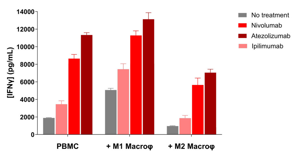
Checkpoint blockade reverses M2-mediated T cell suppression and boosts M1 stimulation

Making a difference as a preclinical CRO in oncology

Why work with Explicyte

Deep immuno-oncology experience

More than 100 in vitro campaigns over ten years, behind 30-plus peer-reviewed publications in immuno-oncology.

See our latest papers →

A dedicated PhD study director

A single PhD-level study director owns your program from experimental plan through final-report discussion.

Learn about our workflow →

Reference-anchored design

Every readout is benchmarked against pathway-inhibitor and checkpoint-antibody controls, so your compound’s effects stay interpretable.

See our publication record →

Mechanism-resolving readouts

IFN-γ plus macrophage cytokine panels, extendable to flow immunophenotyping, proteomics, and transcriptomics for deeper mechanism.

View our platform →

The Explicyte team at their Bordeaux laboratory

Paul Marteau, PharmD (study director), Imane Nafia, PhD (CSO), Loïc Cerf, MSc (COO), Alban Bessède, PhD (founder, CEO), Jean-Philippe Guégan, PhD (CTO)

Contact our team

Discuss your macrophage study

Tell us about your macrophage program — polarization, M2 suppression, or a combination strategy — and we'll propose a fit-for-purpose study.

Answers about macrophage assays

Frequently asked questions

What does the M1/M2 macrophage assay measure?

Whether a test compound shifts macrophage polarization between M1 and M2 states, and how that changes T cell activation, read as IFN-γ, alongside macrophage cytokines (IL-6, IL-10, IL-12).

Monocytes are isolated from healthy-donor PBMCs and differentiated in vitro into M1 or M2 macrophages, then used alone or co-cultured with autologous activated PBMCs or CD4+ T cells.

It co-cultures monocyte-derived M2 macrophages with activated CD4+ T cells (or PBMCs) and quantifies IFN-γ as a surrogate of T cell activation, to test whether a compound relieves M2-mediated suppression.

M1 macrophages boost T cell activation and raise IFN-γ in co-culture; M2 macrophages suppress it and lower IFN-γ. Compounds are scored by how far they move each direction.

Signaling-pathway inhibitors (p38 MAPK, STAT3, PI3K) and checkpoint antibodies: nivolumab (anti-PD-1), atezolizumab (anti-PD-L1), and ipilimumab (anti-CTLA-4).

Yes. Candidates run as single agents or in combination, which suits checkpoint-plus-pathway strategies aimed at repolarizing M2 macrophages or relieving their suppression of T cells.

M2 macrophages share key features with TAMs — checkpoint engagement, anti-inflammatory cytokines, and immunosuppressive metabolism — so the M2 suppression assay is a practical in vitro proxy for TAM-directed compounds.

Extended macrophage and effector-cell cytokine profiling, flow-cytometry immunophenotyping, proteomics, and transcriptomics, depending on the mechanism you need to resolve.

Explicyte Oncology CRO logo

Capabilities

Modalities