Publication in Molecular Cancer

Why advanced gastric cancers resist PD-1 plus anti-angiogenic therapy — and how M2 macrophages mark the non-responders

Explicyte collaborated with: Bayer·Centre Léon Bérard·CHRU de Brest·Inserm·Institut Bergonié·Institut Cancérologie Montpellier·Institut Gustave Roussy·National Cancer Center (Tokyo)·National Cancer Center Hospital East (Japan)·University of Bordeaux
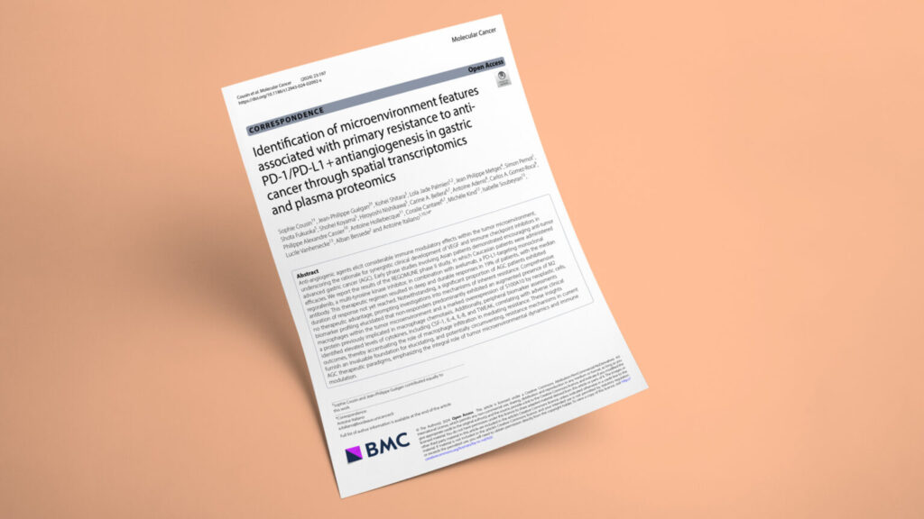
Identification of microenvironment features associated with primary resistance to anti-PD-1/PD-L1 + antiangiogenesis in gastric cancer through spatial transcriptomics and plasma proteomics
GastrointestinalBiomarker analysisBiomarker discoveryDiscoverypre/post studyTrialsFFPE tissuePlasmaBioinformaticsGeoMx DSPMultiplex IF/IHCProteomics
JournalMolecular Cancer
DateSep 2024
Read full paper →

Pairing a VEGFR inhibitor with a PD-1/PD-L1 blocker helps only a fraction of advanced gastric cancer patients — and why the rest resist has stayed unclear. Working from baseline tumor and plasma samples across the REGOMUNE and REGONIVO phase II trials, the team profiled the tumor microenvironment with spatial transcriptomics, multiplex immunofluorescence, and Olink plasma proteomics. Non-responders were defined by M2 macrophage enrichment, tumor-cell S100A10 overexpression, and elevated circulating CSF-1, IL-4, IL-8, and TWEAK — recasting primary resistance as a macrophage-driven process with a candidate blood-based readout.

This study in Molecular Cancer — led by Prof. Antoine Italiano at Institut Bergonié (Bordeaux), with Sophie Cousin and Explicyte’s Jean-Philippe Guégan as co-first authors — dissects why the regorafenib–avelumab combination fails in a large share of advanced gastric cancer (AGC) patients. The regimen produced deep, durable responses in 19% of patients in the REGOMUNE trial, but most saw no benefit, prompting a search for what separates responders from non-responders. Sponsored by Institut Bergonié with funding from Bayer, the work paired clinical outcomes from the REGOMUNE and REGONIVO trials with deep tumor and plasma profiling. Explicyte contributed the spatial transcriptomic profiling, the multiplex immunofluorescence, and the Olink plasma proteomics that anchored the resistance analysis.

The question

Why do some advanced gastric cancers resist PD-1/PD-L1 blockade combined with anti-angiogenic therapy — and what marks the patients who won't respond?

Key steps

  1. 1

    Spatial profiling of responder vs. resistant tumors

    Explicyte ran GeoMx DSP whole-transcriptome atlas profiling on six baseline gastric tumors (three responders, three non-responders), segmenting each ROI into tumor (PanCK+) and immune (CD45+) compartments to map expression of more than 18,000 protein-coding genes. Unsupervised clustering cleanly separated responders from non-responders in the immune compartment, with CD163 overexpressed in resistant patients. Gene Ontology enrichment pointed to macrophage functions, and SpatialDecon deconvolution singled out M2 macrophages as the population most enriched in non-responders.

  2. 2

    Multiplex IHF validation across 43 biopsies

    To validate the signal, Explicyte built two multiplex immunofluorescence panels on the Ventana Discovery platform (Akoya Opal reagents, PhenoImager HT scanning): panel 1 (CD8 / CD11b / CD68 / HLA-DR / PanCK) to distinguish M1 (HLA-DR+) from M2 (HLA-DR−) macrophages, and panel 2 (S100A10 / PanCK). Applied to all available whole-section baseline biopsies from REGOMUNE and REGONIVO (n=43), the panels confirmed M2 enrichment and a higher M2/M1 ratio in non-responders. A high M2/M1 ratio tracked with markedly shorter PFS (2.59 vs. 11.28 months, p=0.005).

  3. 3

    Tumor-cell S100A10 flags poor responders

    Comparative transcriptomics of the tumor compartment showed strong upregulation of S100A10 — a macrophage-chemotaxis protein — alongside its partner Annexin A2 in non-responders. IHF confirmed the pattern at the protein level, with high tumor-cell S100A10 associated with a lower response rate (27.6% vs. 57.1%, p=0.06) and a trend toward shorter PFS (2.72 vs. 7.56 months, p=0.081).

  4. 4

    Plasma proteomics nominates circulating CSF-1

    Explicyte profiled baseline plasma with the Olink Target 96 Immuno-Oncology panel (Proximity Extension Assay). Cytokines tied to macrophage recruitment — CSF-1, IL-4, IL-8, and TWEAK — were elevated in poor-outcome patients and correlated with tissue M2 abundance. CSF-1–high patients fared far worse than CSF-1–low: PFS 1.78 vs. 4.41 months (P<0.001), OS 7.2 vs. 13.48 months (P=0.009), and objective response 0% vs. 43.2% (p=0.003).

Impact

The work reframes primary resistance to checkpoint-plus-antiangiogenic therapy in gastric cancer as a macrophage problem — and surfaces circulating CSF-1 as a candidate blood-based marker of who won't benefit.

19%
objective response rate to regorafenib + avelumab in advanced gastric cancer
0% vs 43.2%
response rate in CSF-1–high vs CSF-1–low patients (p=0.003)
p=0.005
high M2/M1 macrophage ratio linked to shorter PFS (2.59 vs 11.28 months)

For drug developers, the data argue that adding a VEGFR TKI to PD-1/PD-L1 blockade isn’t enough to overcome a macrophage-rich microenvironment — making tumor-associated macrophages, and the CSF-1/CSF-1R axis in particular, a rational co-target for the next generation of gastric cancer combinations. For trial design, plasma CSF-1 offers a peripheral, easily sampled candidate for stratifying likely non-responders up front, especially since PD-L1 CPS showed no predictive value in this setting. Tumor-cell S100A10 adds a second, tissue-based resistance flag worth prospective evaluation.

Profiling resistance to checkpoint or anti-angiogenic combinations? Let's talk spatial, multiplex IHF, and plasma proteomics readouts.

Talk to us
Explicyte Oncology CRO logo

Capabilities

Modalities