
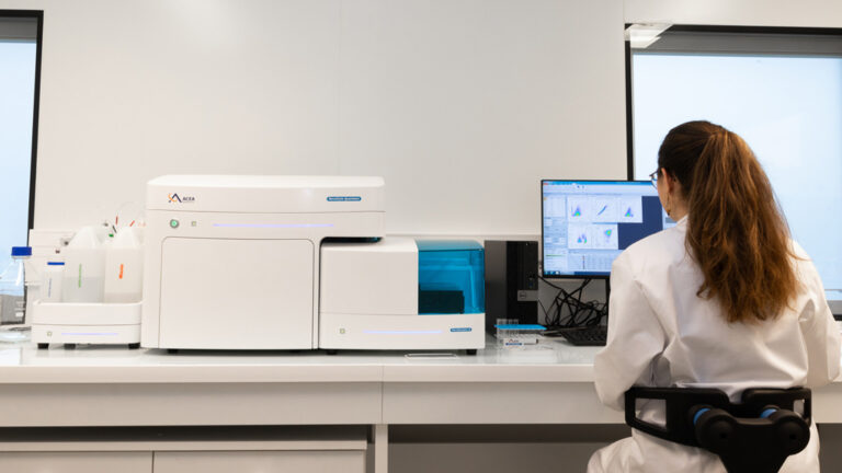
Ascites is a pathological accumulation of peritoneal fluid frequently associated with ovarian cancer. In epithelial ovarian cancer, malignant ascites create a unique tumor microenvironment in which cancer cells can spread and interact with immune cells, non-tumor cells, and soluble mediators.
Ovarian cancer ascites represent a complex mixture of cellular components and soluble factors that can influence tumor cell behavior, immune cell activity, and therapeutic response.
This ascitic microenvironment is increasingly recognized as a pro-inflammatory, immunosuppressive, and tumor-promoting milieu. Although several individual ascites components have been associated with prognosis or chemoresistance, comprehensive profiling is needed to better understand their biological activity.
Study Objective
The objective of this case study was to characterize ovarian cancer ascites using immune cell profiling and soluble mediator analysis, and to evaluate how ascites-associated factors may contribute to an immunosuppressive microenvironment.
This approach aimed to highlight the value of ovarian cancer ascites as a liquid biopsy tool and as a relevant substrate for translational research and therapeutic testing.
Methods
Ovarian cancer ascites samples were analyzed using flow cytometry-based marker expression profiling to characterize the nature of immune cell populations present in the ascitic fluid.
In parallel, soluble mediators were quantified to assess the presence of signaling factors potentially involved in immune suppression, tumor promotion, and therapy resistance.
Functional assays were also performed to investigate the biological effects of acellular ascites fluid and selected ascites-associated factors on healthy monocytes undergoing M1 polarization.
Results
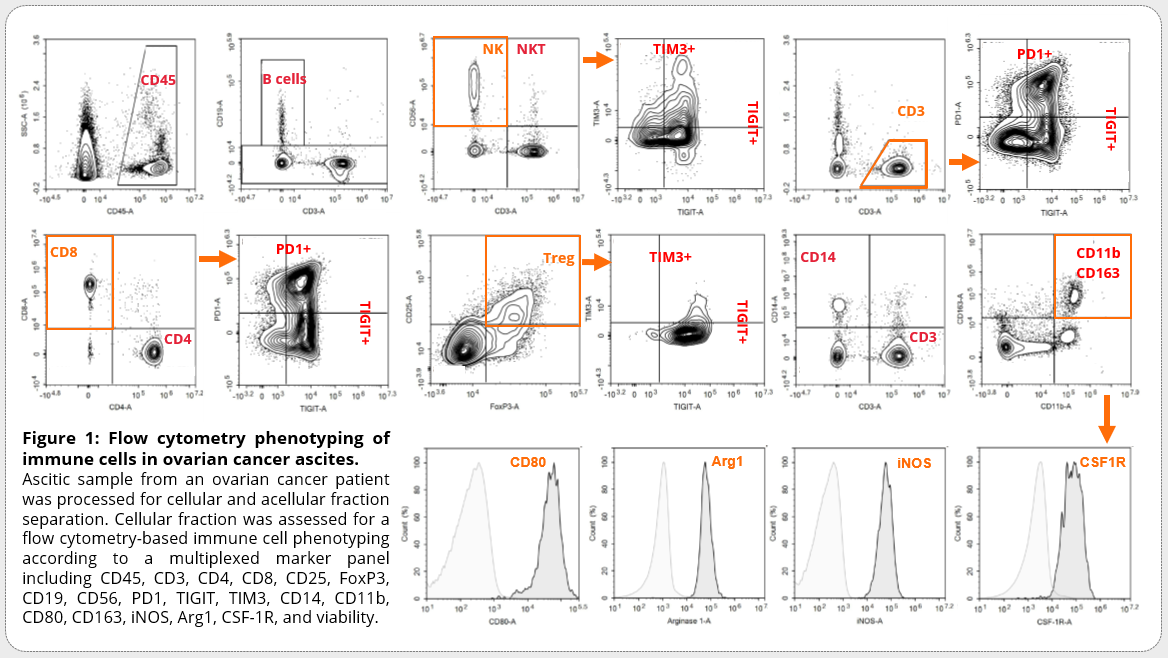
Ovarian cancer ascites were enriched in major immunosuppressive immune cell subsets, including T cell and myeloid populations.
Tumor-associated macrophages displayed a mixed phenotype characterized by elevated CD163 expression, without fitting strictly into classical M1 or M2 macrophage categorization.
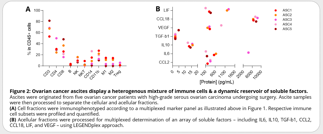
Soluble mediator profiling highlighted a heterogeneous reservoir of increased factors, including IL6, LIF, IL10, and VEGF.
Among these mediators, IL6 and LIF were investigated for their potential role in shaping an immunosuppressive milieu.
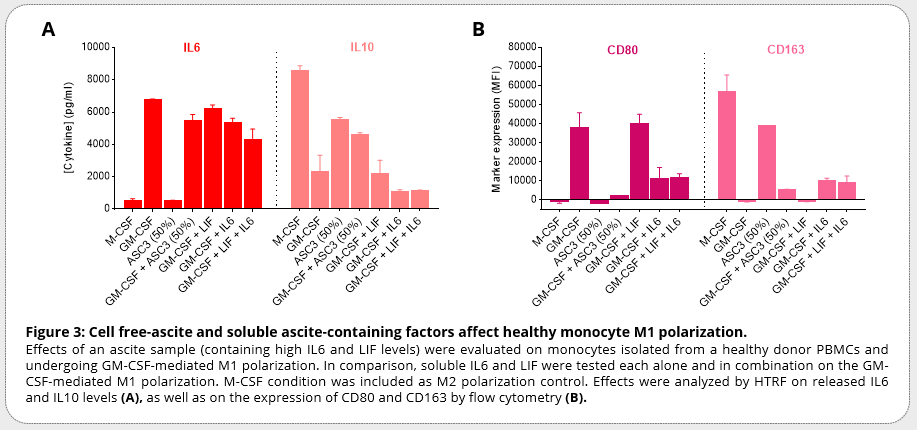
Functional experiments showed that acellular ascites fluid, as well as selected ascites-associated factors, affected healthy monocyte M1 polarization.
LIF alone did not directly affect IL6 or IL10 levels released by the cells. In contrast, IL6 alone or in combination with LIF decreased IL6 levels in the supernatant compared with control conditions, similarly to the effect observed with acellular ascites fluid.
These changes were associated with decreased CD80 expression and increased CD163 expression, supporting a shift toward a more immunosuppressive myeloid phenotype.
Conclusion
This case study highlights the value of ovarian cancer ascites as a biologically relevant material to investigate tumor-immune interactions, suppressive mechanisms, and therapy resistance.
By combining immune cell profiling, soluble mediator quantification, and functional assays, ovarian cancer ascites can provide key insights into the crosstalk between cellular components and soluble factor-mediated signals.
Ovarian cancer ascites therefore represent a remarkable opportunity for translational research, biomarker discovery, therapeutic testing, and the identification of potentially targetable mechanisms involved in immune suppression and tumor progression.