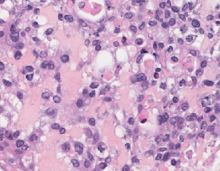
Tumor Organoids & Fragments

Patient-Derived 3D Tumor Models: Immune-Native Organoids and Tumor Fragments

Our team builds 3D tumor models from fresh patient tumor tissues (biopsies or surgical resections), with the native immune compartment preserved.

Reflecting real in-human sensitivity and resistance, they offer an in vitro route to replace in vivo studies and strengthen evidence before trials.

TUMOR ORGANOIDS AND FRAGMENTS

Why immune-native 3D tumor models ?

Patient-derived 3D tumor models are in vitro cultures grown directly from a patient's tumor — as organoids or intact tumor fragments — that preserve both the tumor cells and their native immune populations for functional immunotherapy testing.

Custom 3D models

Through our biobank network, we obtain fresh tumor biopsies for your indication of interest and grow them in 3D at low passage — organoids or intact fragments, built around your program.

Translational relevance

The models preserve tissue architecture, the native tumor microenvironment, and patient heterogeneity. Sensitivity and resistance patterns emerge on human tumor material — reflecting the patient, not a surrogate host.

Mechanistic readouts

Beyond supernatant analysis and kinetic functional readouts, we bring spatial biology — digital pathology and single-cell spatial transcriptomics — to resolve where and how an agent acts, down to mechanism of action.
Imane Nafia, PhD, Chief Scientific Officer at Explicyte

A patient-derived 3D model is only useful if it still contains the patient’s own immune system — ours do. Working in three dimensions also lets us study how a drug penetrates the tumor, and pairing that with spatial platforms shows us where it acts and how the immune response plays out, cell by cell.

Imane Nafia, PhD

Chief Scientific Officer, Explicyte

Early TUMOR organoids

Immune-native tumor organoids (PDTOs)

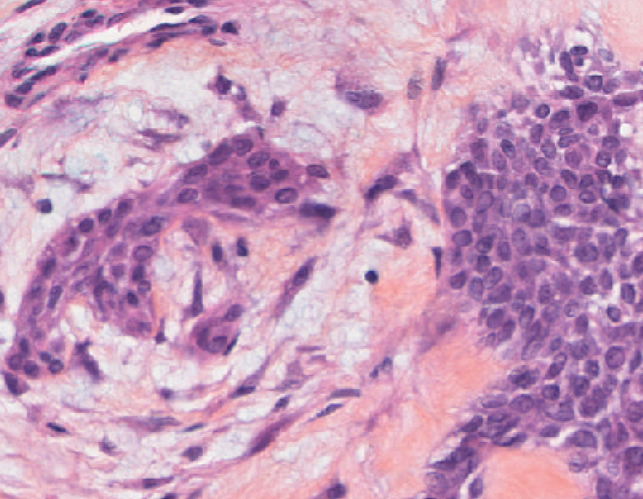
True early-stage organoids derived directly from patient tissue, with no or minimal passage — so the native immune compartment is still present and functional when you test on them.

At a glance

Core features of early PDTOs
Origin
Fresh patient tumor tissues, no/minimal passage
Immune content
Native — CD45, CD3, CD4, CD8 identifiable
Readouts
Flow immune profiling, treatment-response kinetics
Timeline
Short (early / ~1-week window)
Best for
Early immunotherapy testing on patient-proximal organoids
Organoid — 1 week Organoid — 1 week
TNBC patient tumor TNBC patient tumor

Preserved immune fraction, IL2-sustained CD8 persistence

Early immune-native TNBC organoids retain a fraction of T cells relative to the parent tumor. Under IL2, the CD8 compartment persists through day 7, where the untreated condition declines — native immune dynamics that respond to culture conditions rather than a static endpoint.
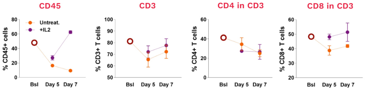
Early immune-native TNBC PDOs display a preserved fraction of T cells, with CD8 IL2-sustained persistence

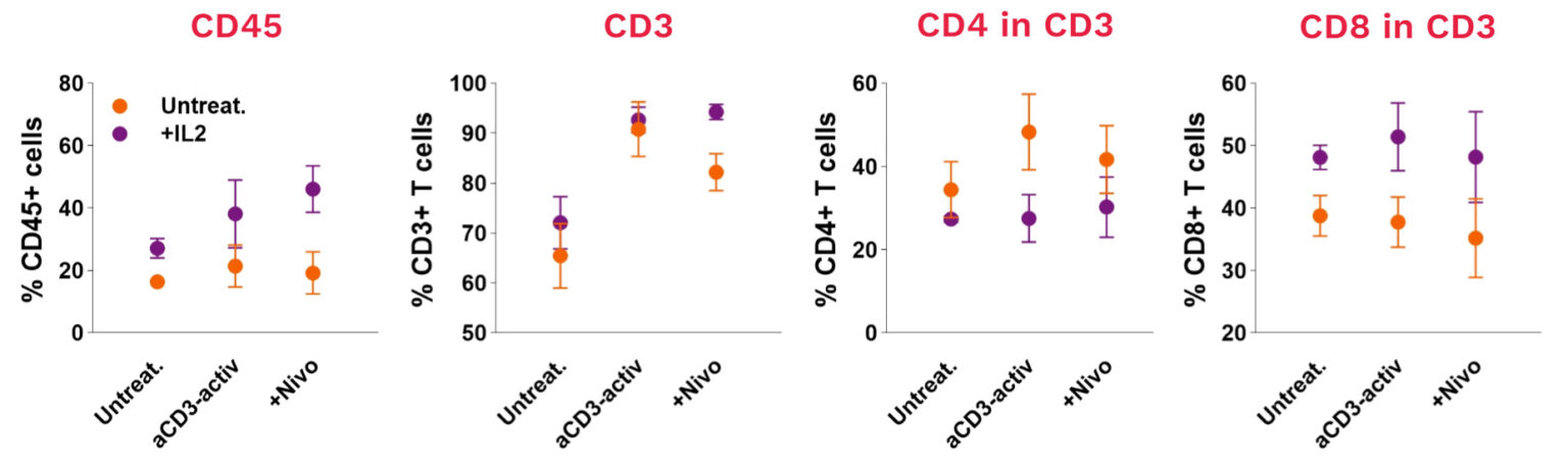
Early immune-native TNBC PDOs are immunotherapy-responsive

The same organoids respond to immunotherapy in vitro: CD3 activation and nivolumab shift the CD45, CD3, CD4, and CD8 compartments, giving a functional readout of immune engagement on patient-proximal material.

Early immune-native TNBC PDOs are immunotherapy-responsive

PDTF MODELS

Patient-Derived Tumor Fragments

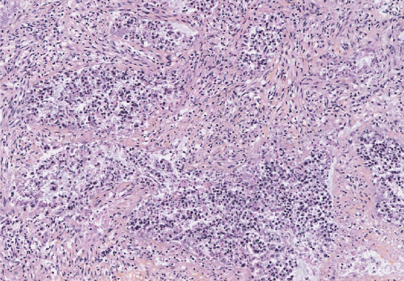
The closest 3D model to the patient tumor — intact fragments that recapitulate tissue features and architecture, with the native immune infiltrate preserved in place.

At a glance

Core features of PDTFs

Origin
Fresh patient tumor tissues, tissue architecture retained
Immune content
Native infiltrate in place — CD45, CD8, myeloid (CD11b/c), CD16, CD19
Readouts
Flow immune profiling, IFN-γ / secretome, treatment-response
Timeline
Short (2–5 day windows)
Best for
Immunotherapy response and biomarker evaluation on intact tumor tissue before clinical trial initiation
2-day CRC fragment under aCD3 treatment

2-day colorectal cancer fragment, treated with anti-CD3

A quantifiable native immune infiltrate

Flow cytometry of a CRC fragment resolves the native immune infiltrate directly from patient tissue — T cells (CD8, CD4), a dominant myeloid compartment (CD11b, CD11c), NK (CD16), and B cells (CD19) — each measurable as a fraction of viable CD45⁺ cells before any treatment.
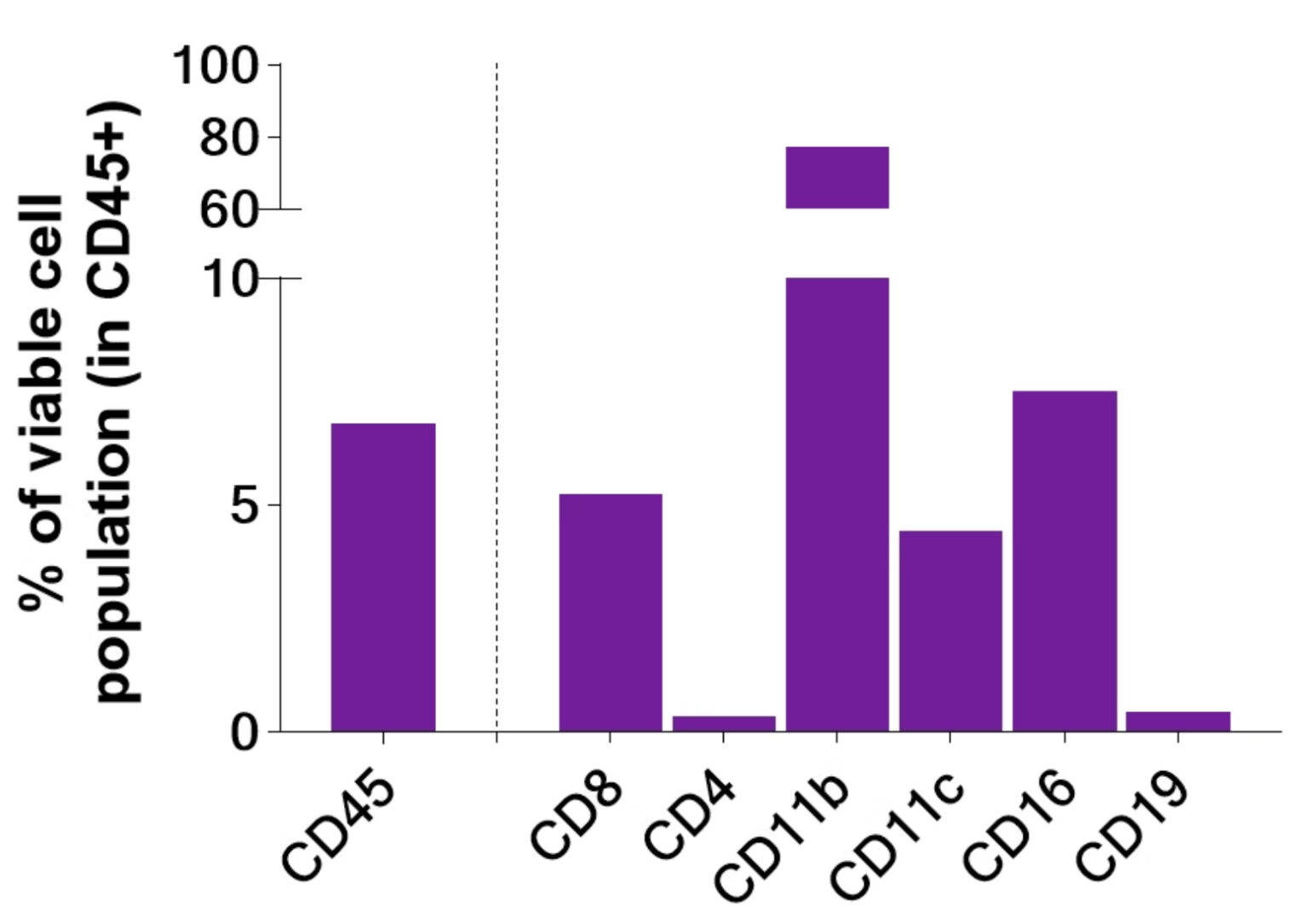
tumor immune infiltration CRC tumor fragment

Immune subsets modulated under immunotherapy

Native immune cells in CRC fragments shift under treatment. Across 2- and 5-day windows, aCD3 and aCD3 + nivolumab modulate the CD45, CD8, CD11b, and CD11c compartments — the native infiltrate responding, not just persisting.
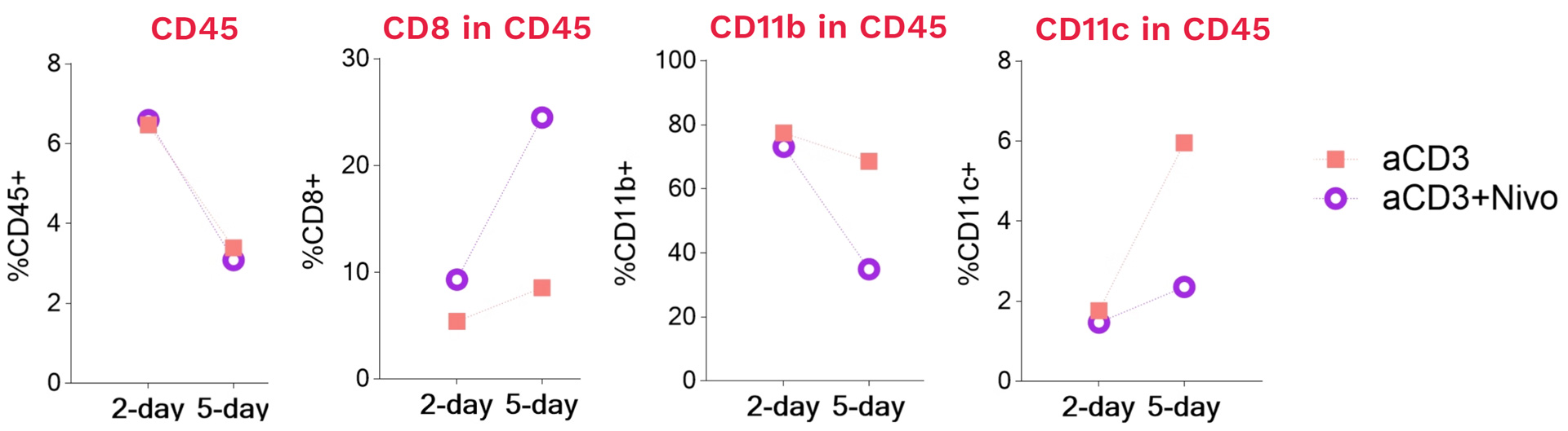
Native immune cells in CRC tumor fragments are modulated under immunotherapy

Functional IFN-γ response

Fragments are functionally responsive to immunotherapy: aCD3 + nivolumab drives a marked IFN-γ release over untreated and single-agent conditions at both 2 and 5 days — a functional secretome readout on intact patient tissue.
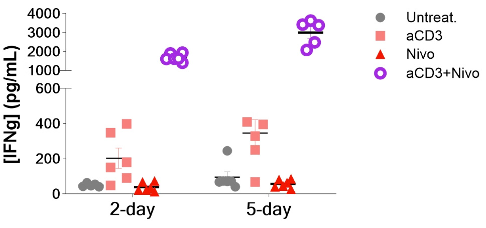
Immune-native CRC fragments are functionally responsive to immunotherapy

Making a difference as a preclinical CRO in oncology

Why work with Explicyte

Proven experience

>10 years in preclinical immuno-oncology, with deep expertise in functional assays and tumor microenvironment profiling across in vitro and ex vivo models.

More about us →

Built around your program

A PhD-level study director designs your study around your indication, tumor model, and immunotherapy, sourcing fresh biopsies through our biobank network.

Learn about our workflow →

Beyond functional readouts

A multiomic platform to resolve mechanism of action alongside the functional data — flow cytometry, secretome analysis, digital pathology, and single-cell spatial transcriptomics.

View our platform →

Reports you can act on

Progress reports let you adjust the plan as the study runs; final reports arrive with ready-to-publish data that holds up in due diligence.

See our publication record →

The Explicyte team at their Bordeaux laboratory

Paul Marteau, PharmD (study director), Imane Nafia, PhD (CSO), Loïc Cerf, MSc (COO), Alban Bessede, PhD (founder, CEO), Jean-Philippe Guégan, PhD (CTO)

Contact our team

Discuss your 3D tumor model study

Tell us about your indication, the immunotherapy you're testing, and the questions you want the model to answer — we'll scope the right model (organoids or fragments), readouts, and reporting, and send a fit-for-purpose proposal.

Patient-derived 3D tumor models: common questions

Frequently asked questions

What are patient-derived 3D tumor models?

They are in vitro cultures grown directly from a patient’s tumor that preserve both the tumor cells and their native immune populations. Explicyte offers two formats: immune-native organoids (PDOs), grown from patient tissue at no or minimal passage, and patient-derived tumor fragments, which keep the tissue architecture and immune infiltrate intact. Both allow functional immunotherapy testing on patient-proximal material, in vitro.

Organoids are early-stage cultures grown from patient tissue with no or minimal passage, suited to short-timeline immunotherapy testing where a preserved immune fraction is enough. Tumor fragments are the closest model to the patient tumor: they retain intact tissue architecture, the native tumor microenvironment, and the immune infiltrate in place, making them well suited to studying immune-cell modulation and functional response on genuine tissue. We help you choose the format that fits your program and question.

Yes. Native immune populations survive into culture and stay identifiable and functional — including T cells (CD3, CD4, CD8), myeloid cells (CD11b, CD11c), NK cells (CD16), and B cells (CD19), quantified by flow cytometry as a fraction of viable CD45⁺ cells. In organoids, the CD8 compartment can be sustained under IL2; in fragments, the native infiltrate is modulated under immunotherapy rather than simply persisting.

The models respond to a range of immunotherapy approaches in vitro, including checkpoint inhibitors such as anti-PD-1 (nivolumab), T-cell–activating agents such as anti-CD3, and combinations. Each study is configured to the mechanism the therapeutic depends on. Contact us to discuss whether your modality fits the model.

Readouts span three layers: flow-cytometry immune profiling of native populations, functional secretome analysis of the supernatant including kinetic cytokine readouts such as IFN-γ, and spatial biology — digital pathology and single-cell spatial transcriptomics — to resolve where and how an agent acts, down to mechanism of action.

They offer an in vitro alternative to animal models that keeps what animal models cannot: intact human tissue architecture, the native tumor microenvironment, and real patient heterogeneity. Sensitivity and resistance patterns emerge on human tumor material rather than in a surrogate host, which can reduce reliance on in vivo work and strengthen evidence ahead of trials.

Through our biobank network, we obtain fresh tumor biopsies matched to your indication of interest. We have generated immune-native models from indications including triple-negative breast cancer (TNBC) and colorectal cancer (CRC), and can scope other solid-tumor indications on request.

Explicyte Oncology CRO logo

Capabilities

Modalities