Publication in Science Advances

How normal-appearing vitiligo skin keeps its pigment: PD-1 and regulatory T cells restrain disease-ready CD8 T cells

Explicyte collaborated with: University of Bordeaux
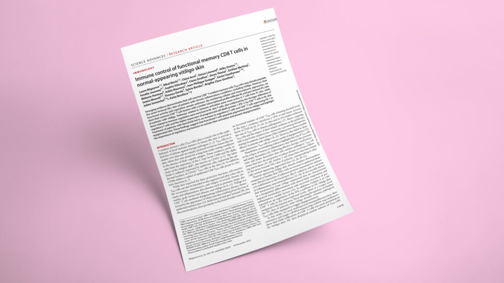
Immune control of functional memory CD8 T cells in normal-appearing vitiligo skin
JournalScience Advances
DateDec 2025
Read full paper →

In vitiligo, white patches form when CD8 T cells destroy melanocytes — yet a patient's normal-appearing, fully pigmented skin already harbors the same pathogenic T cell clones. So why does it keep its color? Using single-cell RNA-seq, TCR sequencing, multiplex immunofluorescence, and flow cytometry on paired skin biopsies, the team found that never-lesional skin is enriched for regulatory T cells and PD-1–high CD8 T cells that hold these disease-ready cells in check — and that effective therapy strengthens that control.

Published in Science Advances, this study from the ImmunoConcEpT laboratory (CNRS UMR 5164, University of Bordeaux) and the Department of Dermatology at Bordeaux University Hospital — led by Katia Boniface and Julien Seneschal, with Laure Migayron as first author — maps the immune landscape of clinically normal-appearing skin in vitiligo. Explicyte contributed the multiplex immunofluorescence and multispectral imaging (Ventana Discovery, Akoya Opal, PhenoImager HT) that resolved CD8 T cell subsets, regulatory T cell infiltration, and PD-1/PD-L1 spatial relationships across never-lesional, peri-lesional, and treated skin. Several of those analyses drew on biopsies from the phase 2 BARVIT trial (NCT04822584) of baricitinib plus phototherapy, where Explicyte performed paired pre/post immune profiling.

The question

If the normal-appearing skin of vitiligo patients is already populated by functional, disease-capable CD8 T cells, why doesn't it lose its pigment?

Key steps

  1. 1

    Single-cell mapping of paired vitiligo skin

    scRNA-seq on the 10x Genomics Chromium platform profiled never-lesional (NL) and peri-lesional (PL) skin from six patients with active vitiligo — a final dataset of 77,532 cells across 12 skin cell types and 17 T cell clusters. Both sites carried the same CD8 T cell clusters and resident-memory subsets; PL skin simply held more T cells. Matched NL and PL samples were transcriptionally similar across all six patients.

  2. 2

    Same T cell repertoire across both sites

    High-throughput TCRβ sequencing in eight patients (plus four healthy-skin controls) showed NL and PL skin share many of the same polyclonal T cell clones, distinct from healthy skin and with no dominant emergent clone. The normal-looking skin is already seeded with the same disease-associated repertoire as the lesion border.

  3. 3

    Multiplex IF phenotyping of CD8 TRM subsets

    Explicyte’s multispectral imaging (Ventana Discovery + Akoya Opal panels on the PhenoImager HT, MelanA/CD8/CD69/CD103) and multiparametric flow cytometry quantified resident-memory subsets in paired skin from six to seven patients. NL and PL skin carried similar CD69/CD103-defined CD8 TRM subsets, and CD8 T cells were elevated in both versus healthy skin — confirming that the regulatory difference lies in cell state, not subset identity.

  4. 4

    Primed to inflame, but held by regulatory brakes

    Ex vivo anti-CD3 (OKT-3) activation of NL skin explants drove a vitiligo-like inflammatory program (CXCL9, CXCL10, TNF, STAT1, MX1); NanoString profiling of 579 immune genes found 125 DEGs versus healthy skin but zero between NL and PL skin — NL skin responds essentially like PL once triggered. CellChat on the scRNA-seq data then showed NL skin enriched for 21 immune-regulatory signaling pathways (CD96, TGFβ, CD39/ENTPD1) versus 11 activation pathways in PL, alongside more regulatory T cells and a higher FOXP3/CD8 ratio.

  5. 5

    PD-1 restrains the cells — and therapy reinforces it

    Flow cytometry and Explicyte’s multispectral imaging showed higher PD-1 on NL CD8 T cells and more PD-L1+ CD11c+ dendritic cells in NL skin; PD-1 blockade with nivolumab on NL explants increased IFN-γ, confirming PD-1 actively suppresses these cells. In paired BARVIT-trial biopsies (six patients, baseline vs 9 months), the same multiplex IF workflow showed reduced CD8 infiltration plus increased PD-1+ CD8 T cells, PD-L1+ DCs, and a higher FOXP3/CD8 ratio in repigmented skin.

Impact

The work reframes vitiligo's normal-looking skin as an actively policed pre-lesional state: the disease-driving T cells are already present, but regulatory T cells and PD-1/PD-L1 signaling keep them quiet. That makes the brakes themselves — not just the effector cells — a target worth engaging.

77,532
single cells profiled across paired never-lesional and peri-lesional vitiligo skin
0
differentially expressed genes between never-lesional and peri-lesional skin after T cell activation (vs 125 vs healthy skin)
21
immune-regulatory signaling pathways enriched in never-lesional skin (vs 11 activation pathways in peri-lesional skin)
9 months
of baricitinib + phototherapy raised PD-1⁺ CD8 T cells and the FOXP3/CD8 ratio in repigmented skin

For drug developers, durable vitiligo control may depend less on eliminating resident memory T cells — which JAK inhibitors fail to clear — and more on sustaining the regulatory checkpoints that keep those cells dormant. Agents that engage PD-1/PD-L1 or expand regulatory T cell activity become rational strategies, and the data offer a mechanistic explanation for why anti–PD-1 cancer therapy can trigger vitiligo. The same single-cell-plus-multiplex-imaging workflow translates directly to mapping checkpoint and regulatory biology in tumor tissue.

Mapping checkpoint and regulatory T cell biology in patient tissue? Let's talk multiplex imaging and single-cell study design.

Talk to us
Explicyte Oncology CRO logo

Capabilities

Modalities