
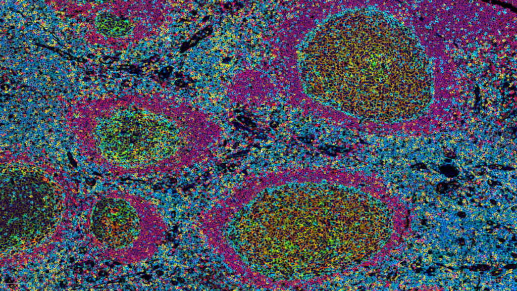
B cells are increasingly recognized as key contributors to anti-tumor immune responses and response to cancer immunotherapy. Recent studies have demonstrated the association between B cell-enriched immune signatures and response to immunotherapy across several cancer indications, including melanoma, renal cell carcinoma, and sarcoma.
In responding patients, B cells are frequently enriched within organized lymphoid aggregates known as tertiary lymphoid structures, or TLS. Mature TLS, particularly those containing germinal center-like features, support B cell development, antigen presentation, and immune cell interactions that may contribute to the initiation and regulation of anti-tumor immunity.
The presence of TLS before treatment initiation has also been associated with improved clinical outcome, placing B cells and TLS at the forefront of cancer immunotherapy research.
Study objective
The objective of this case study was to establish a histological and quantitative workflow to characterize TLS in human tumor samples.
This approach was designed to support B cell and TLS-focused research in cancer immunotherapy by enabling:
- identification of TLS within tumor tissue sections;
- characterization of TLS immune composition;
- distinction between immature and mature TLS-associated B cell populations;
- quantitative assessment of TLS size, density, and spatial organization.
Analytical approach
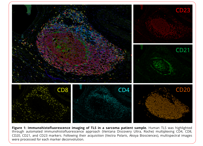
Explicyte developed a TLS analysis workflow based on automated staining, slide digitization, and quantitative image analysis.
The workflow combines multiparametric immunohistofluorescence with digital pathology to characterize immune cell populations within TLS and their spatial organization in the tumor microenvironment.
The staining panel includes key immune and TLS-associated markers:
- CD4, for helper T cells;
- CD8, for cytotoxic T cells;
- CD20, for B cells;
- CD21, for follicular dendritic cells;
- CD23, to further characterize B cell maturation and TLS organization.
Customized panels and image analysis workflows can also be developed to address specific biological questions or project requirements.
Digital pathology workflow
Tumor tissue sections are stained using an automated multiparametric immunohistofluorescence workflow.
Following staining, slides are digitized and analyzed using image analysis tools to identify and segment TLS regions. Quantitative data can then be generated on TLS-associated features, including:
- TLS density;
- TLS size;
- mature versus immature TLS status;
- immune cell density within TLS;
- spatial mapping of immune cell populations.
This approach allows the generation of reproducible, quantitative, and spatially resolved data from tumor tissue sections.
Results
TLS identification by tissue segmentation
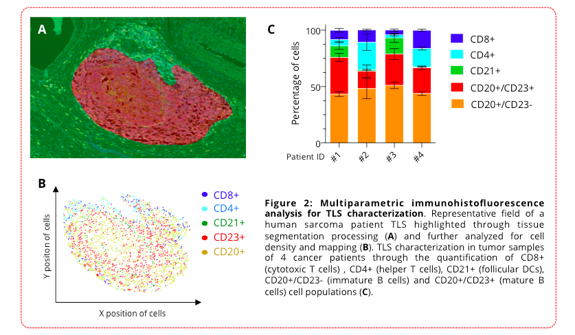
Representative TLS regions were identified in human sarcoma tumor tissue using tissue segmentation processing.
This enabled precise localization of organized lymphoid aggregates within the tumor section and provided the basis for downstream quantitative analysis.
Spatial mapping of TLS immune composition
Following TLS segmentation, immune cell populations were mapped within the TLS area.
This analysis enabled visualization and quantification of the spatial distribution of key immune cell subsets, including CD4+ T cells, CD8+ T cells, CD20+ B cells, and CD21+ follicular dendritic cells.
Quantitative characterization of TLS across tumor samples
TLS were characterized in tumor samples from four cancer patients using quantitative image analysis.
The analysis included the quantification of:
- CD8+ cytotoxic T cells;
- CD4+ helper T cells;
- CD21+ follicular dendritic cells;
- CD20+/CD23− B cells;
- CD20+/CD23+ B cells.
This multiparametric analysis supports a deeper understanding of TLS biology and allows comparison of TLS composition and maturation status across patient samples.
Conclusion
This case study illustrates Explicyte’s ability to characterize B cell-rich tertiary lymphoid structures in human tumor samples using multiplex immunohistofluorescence and quantitative digital pathology.
By combining automated staining, whole-slide imaging, TLS segmentation, and immune cell mapping, this workflow provides a robust platform to investigate the role of B cells and TLS in anti-tumor immune responses.
This approach can support translational research programs focused on immunotherapy response, biomarker discovery, and the characterization of the tumor immune microenvironment.
References
Jonsson et al., Nature, 2020; 577:561–565.
Wargo et al., Nature, 2020; 577:549–555.
Friedman et al., Nature, 2020; 577:556–560.
Bruno, Nature, 2020; 577:474–476.