

Cytokine release is a key functional readout for assessing immune cell activation, modulation, and effector function in immuno-oncology.
While phenotypic analysis provides valuable information on immune cell identity and activation status, the measurement of cytokines released by immune cells offers a direct functional surrogate of their biological activity. Depending on the assay context, cytokine profiling can be used to evaluate T cell activation, NK cell cytotoxicity, macrophage-mediated immunosuppression, or the ability of therapeutic candidates to enhance or inhibit immune responses.
Explicyte has developed a broad in vitro immuno-oncology assay platform combining cell-based models, quantitative cytokine measurement, and high-throughput-compatible readouts to support the functional evaluation of immunotherapeutic candidates.
Study objective
The objective of this case study was to illustrate how cytokine release can be used as a relevant surrogate marker of immune cell subset function across several in vitro immuno-oncology assays.
The platform was designed to evaluate the modulatory potential of candidate therapeutics on immune cell activity using quantitative cytokine readouts, including IFNγ, IL-2, TNFα, IL-6, and IL-10.
Assay principle
Explicyte’s in vitro immune cell-based assays are based on the activation, co-culture, or functional modulation of defined immune cell populations, followed by quantitative measurement of cytokines released into the culture supernatant.
Depending on the biological question, cytokine release can be used to assess:
- PBMC activation upon T cell receptor stimulation;
- T cell activation in mixed lymphocyte reaction assays;
- NK cell activation and antibody-dependent cell-mediated cytotoxicity;
- macrophage polarization and immunosuppressive function;
- pharmacological modulation by immune checkpoint inhibitors or pathway inhibitors.
Cytokines are quantified using sensitive immunoassay-based technologies, including HTRF-compatible readouts, allowing robust and quantitative analysis of immune cell function.
Experimental models and results
PBMC cytokine release upon CD3 activation and immune checkpoint inhibition
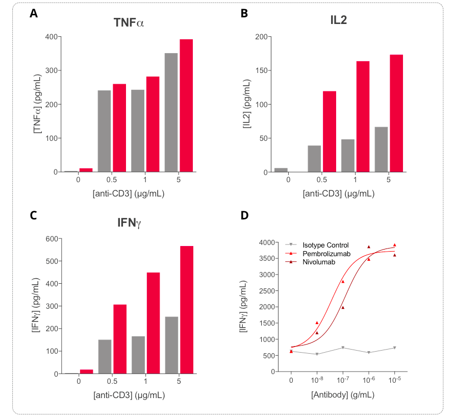
Human PBMCs were stimulated with increasing concentrations of anti-CD3 antibody to induce T cell activation.
Key inflammatory cytokines, including TNFα, IL-2, and IFNγ, were differentially modulated following anti-CD3 stimulation.
TNFα secretion was efficiently induced at 24 hours, while IFNγ and IL-2 release increased in a time- and dose-dependent manner following anti-CD3 treatment.
These results demonstrate that cytokine quantification, particularly IL-2 and IFNγ measurement, provides a valuable functional readout to assess PBMC activation.
In addition, treatment of anti-CD3-activated PBMCs with increasing concentrations of anti-PD-1 antibodies, including Nivolumab and Pembrolizumab, induced a dose-dependent increase in IFNγ release. This confirmed the ability of the assay to detect further optimization of PBMC activation by immune checkpoint blockade.
IFNγ release in a human allogeneic mixed lymphocyte reaction assay
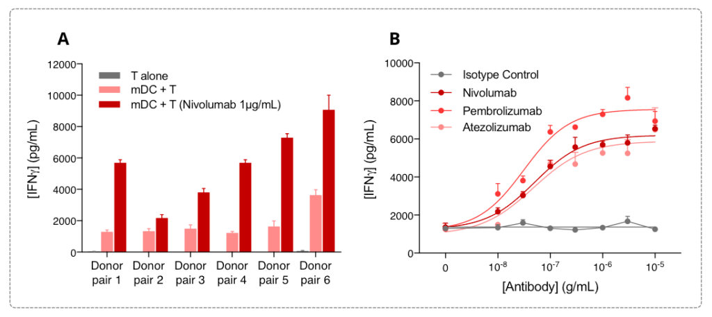
Explicyte’s mixed lymphocyte reaction assay is based on the co-culture of human CD4+ T cells as responder cells with monocyte-derived dendritic cells as stimulator cells.
In this model, IFNγ release provides a functional surrogate of dendritic cell-mediated T cell activation and can be used to evaluate immunotherapeutics designed to enhance T cell responses.
Peripheral blood monocytes from six human donors were differentiated into monocyte-derived dendritic cells and co-cultured with CD4+ T cells isolated from different donors.
Although the amplitude of IFNγ release varied between donor pairs, the MLR response was robust and reproducible. PD-1/PD-L1 blockade further enhanced the response, as shown by dose-dependent increases in IFNγ release following treatment with anti-PD-1 antibodies, including Nivolumab and Pembrolizumab, or the anti-PD-L1 antibody Atezolizumab.
NK cell IFNγ release and antibody-dependent cytotoxicity
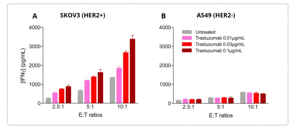
In NK cell-mediated killing assays, IFNγ release is used as a functional marker of NK cell activation and cytotoxic activity.
This readout can support the evaluation of therapeutic candidates that enhance NK cell killing, as well as antibodies targeting tumor antigens and designed to elicit antibody-dependent cell-mediated cytotoxicity.
SKOV3 ovarian tumor cells, which express HER2, and A549 lung tumor cells, which do not express HER2, were cultured under control conditions or treated with increasing doses of Trastuzumab. IL-2-activated primary NK cells were then added at different effector-to-target ratios.
After 24 hours, supernatants were collected and analyzed for IFNγ release.
IFNγ levels increased in an NK ratio-dependent manner in both SKOV3 and A549 co-cultures, reflecting basal NK cell cytotoxic activity. However, Trastuzumab further enhanced IFNγ release only in HER2-positive SKOV3 tumor cells, in both a dose-dependent and NK ratio-dependent manner.
These results demonstrate the ability of the assay to capture tumor antigen-dependent ADCC activity.
Cytokine profiling of M2 macrophage polarization and immunosuppressive function
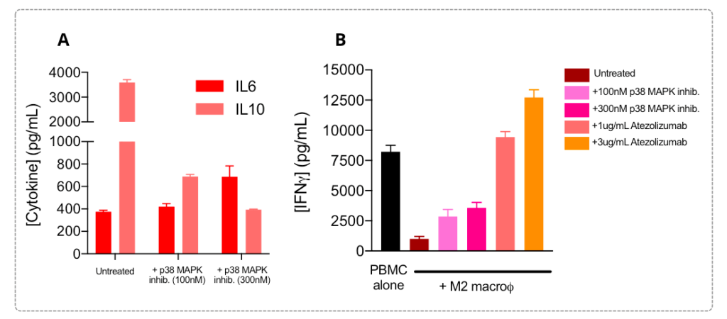
M2 macrophages are characterized by an IL-6low / IL-10high cytokine profile and are known to contribute to T cell response suppression. This phenotype is supported by signaling pathways such as JAK/STAT and SMAD/p38 MAPK.
During M2 macrophage polarization, the addition of a p38 MAPK inhibitor partially and dose-dependently reversed the IL-6low / IL-10high profile, indicating a repolarization or phenotype-switching effect.
In a PBMC co-culture immunosuppression assay, M2 macrophages strongly suppressed T cell activity, as shown by decreased IFNγ release.
When M2 macrophages were treated with a p38 MAPK inhibitor during polarization, their suppressive effect on activated PBMCs was partially reversed in a dose-dependent manner.
Atezolizumab treatment also relieved the immunosuppressive function of M2 macrophages on T cell response, resulting in restoration of IFNγ release.
Conclusion
This case study demonstrates the value of cytokine release as a functional and quantitative readout across multiple in vitro immuno-oncology assay platforms.
By measuring cytokines such as IFNγ, IL-2, TNFα, IL-6, and IL-10, Explicyte’s assays enable the functional characterization of immune cell activation, cytotoxicity, polarization, and immunosuppressive activity.
This platform supports the preclinical evaluation of immunotherapeutic candidates, including immune checkpoint inhibitors, tumor-targeting antibodies, pathway modulators, and combination strategies designed to enhance anti-tumor immune responses.