
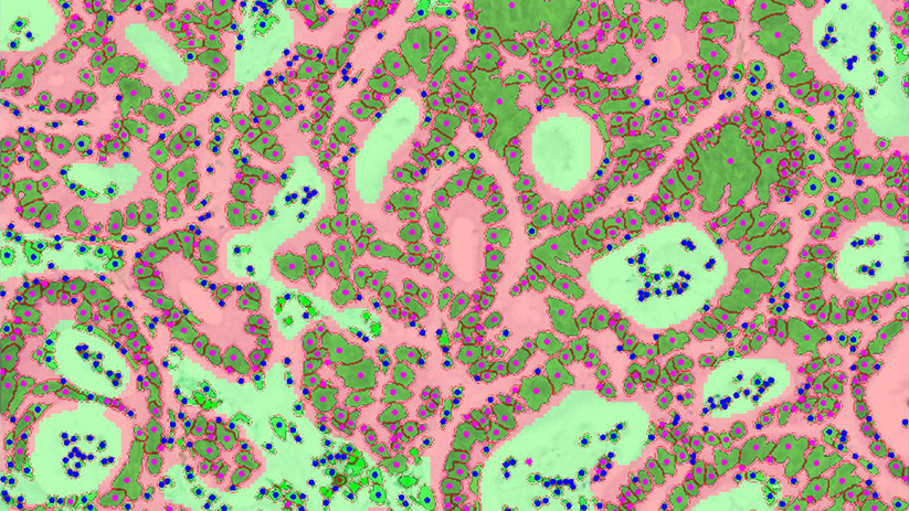
The tumor microenvironment plays a central role in cancer progression and response to immunotherapy. Immune checkpoint inhibitors targeting the PD-1/PD-L1 axis and CTLA-4 have demonstrated major clinical benefit across several cancer indications, highlighting the importance of the tumor immune landscape in shaping therapeutic response.
However, response to immune checkpoint blockade remains heterogeneous and depends on multiple parameters, including the density, localization, and spatial organization of immune cells within the tumor microenvironment.
To address this challenge, Explicyte has developed an integrated quantitative histology and digital pathology platform to characterize the tumor immune contexture in FFPE tissue samples.
Study objective
The objective of this case study was to quantitatively characterize the immune contexture of human lung adenocarcinoma samples using multiplex immunohistochemistry, multispectral imaging, and image analysis.
The workflow was designed to evaluate:
- tumor and stromal tissue compartments;
- immune cell density within each region;
- immune cell phenotypes;
- spatial proximity between immune cells and tumor cells;
- inter-patient heterogeneity in immune infiltration patterns.
Analytical approach
Explicyte implemented a fully integrated workflow spanning from FFPE slide preparation and automated staining to image digitization and computational analysis.
The approach can be applied in monoplex or multiplex format using conventional immunohistochemistry or immunohistofluorescence, depending on the project objectives and biomarker panel.
In this case study, multiplex immunohistochemistry was performed on human FFPE lung adenocarcinoma sections using a panel combining:
- CK7, to identify tumor cells;
- CD8, to identify cytotoxic T cells;
- CD163, to identify tumor-associated macrophages.
Slides were stained on an automated Ventana Discovery platform, acquired using a multispectral imaging system, and analyzed using image analysis software supported by machine-learning-based tissue and cell segmentation.
Digital pathology workflow
The image analysis workflow included several key steps:
- First, tissue segmentation was performed to distinguish tumor and stromal regions.
- Second, cell segmentation and phenotyping were applied to identify tumor cells and immune cell populations based on marker expression.
- Finally, quantitative spatial analyses were performed to measure immune cell density and proximity of immune cells to tumor cells.
This approach generated quantitative data on both immune cell infiltration and spatial organization within the tumor microenvironment.
Results
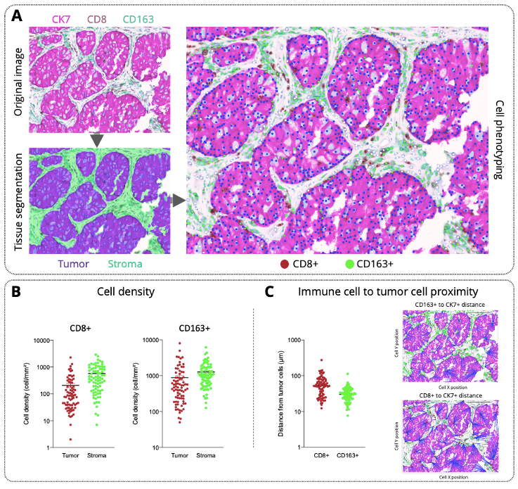
Multiplex staining enables simultaneous visualization of tumor and immune populations
Human lung adenocarcinoma FFPE sections were stained using a multiplex panel combining CK7, CD8, and CD163.
This enabled simultaneous visualization of tumor cells, cytotoxic T cells, and tumor-associated macrophages within the same tissue section.
The acquired images were processed to segment tumor and stromal compartments and to identify individual cell phenotypes within each region.
Immune cell density differs between tumor and stromal regions
Quantitative analysis of 80 patient samples showed that CD8+ T cells and CD163+ macrophages were detected across both tumor and stromal compartments.
Immune cell density was higher in the stroma compared with tumor regions, highlighting the importance of spatial compartmentalization when evaluating tumor immune infiltration.
The analysis also revealed a heterogeneous immune infiltration profile across patients, which may contribute to differences in sensitivity or resistance to immunotherapies.
Spatial proximity analysis reveals patient-dependent immune organization
Beyond immune cell density, the proximity of CD8+ T cells and CD163+ macrophages to CK7+ tumor cells was calculated for each patient sample.
This spatial analysis demonstrated patient-dependent patterns of immune cell localization relative to tumor cells.
Such information provides deeper insight into the organization of the tumor immune microenvironment and may help identify spatial biomarkers associated with therapeutic response.
Conclusion
This case study illustrates Explicyte’s ability to characterize the tumor immune contexture using quantitative histology, multiplex immunohistochemistry, multispectral imaging, and digital pathology analysis.
By combining automated staining, tissue segmentation, cell phenotyping, immune cell density quantification, and spatial proximity analysis, the platform provides a robust approach to investigate the immune landscape of human tumor samples.
This workflow can support translational research programs, biomarker discovery, and mechanism-of-action studies for innovative cancer immunotherapies.