
Background
Most cancer patients treated with anti-PD1 or anti-PDL1 immune checkpoint blockers do not derive clinical benefit. There is therefore a crucial need to identify reliable predictive biomarkers of response.
Beyond PD1, several immune checkpoints, including CTLA4, LAG3, TIM3 and TIGIT, are associated with a T cell exhausted phenotype and play an important role in cancer immune evasion.
However, the impact of simultaneous expression of multiple inhibitory receptors by T cells on clinical outcome in patients treated with immune checkpoint blockers remains poorly understood.
Methods
Tumor tissue samples collected before immune checkpoint blocker initiation were analyzed from patients with solid tumors included in an institutional molecular profiling program.
Multiplex immunohistofluorescence was performed using a panel combining CD3, PD1, TIM3, LAG3, TIGIT and DAPI.
Immune cell characterization was then performed using multispectral image analysis and digital pathology workflows.
The association between co-expression of T cell-associated exhaustion markers, clinical response rate, progression-free survival and overall survival was investigated using Cox proportional hazards models.
Results
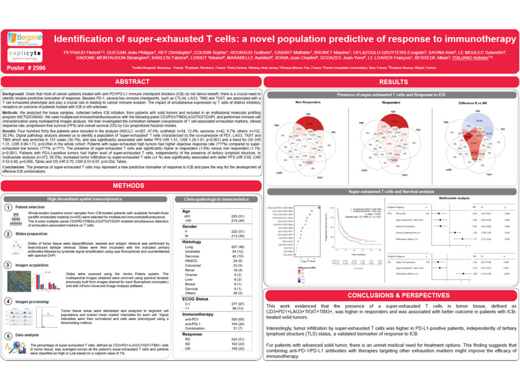
A total of 435 patients were included in the analysis, including patients with non-small cell lung cancer, urothelial carcinoma, sarcoma and other solid tumors.
Digital pathology analysis identified a population of “super-exhausted” T cells characterized by the co-expression of PD1, LAG3, TIGIT and TIM3.
This super-exhausted T cell population was enriched in 131 cases, corresponding to 30.1% of the cohort.
In the whole cohort, the presence of super-exhausted T cells was significantly associated with improved progression-free survival and showed a trend toward improved overall survival.
Patients with tumors enriched in super-exhausted T cells showed a higher objective response rate compared with patients with low levels of super-exhausted T cells.
Super-exhausted T cells were significantly more abundant in responders compared with non-responders.
Patients with PD-L1-positive tumors had higher levels of super-exhausted T cells, independently of the presence of tertiary lymphoid structures.
In multivariate analysis, increased tumor infiltration by super-exhausted T cells, defined as 1% or higher, was significantly associated with better progression-free survival and overall survival.
Conclusion
These findings suggest that the presence of super-exhausted T cells may represent a new predictive biomarker of response to immune checkpoint blockers.
The co-expression of multiple inhibitory receptors on T cells may identify a biologically relevant immune cell population associated with improved clinical benefit.
This work also supports the potential development of rational immune checkpoint blocker combinations targeting complementary exhaustion pathways.