
Background
Immune checkpoint inhibitors have revolutionized the management of patients with non-small cell lung cancer. However, most patients with advanced NSCLC display primary resistance to these treatments.
Indoleamine 2,3-dioxygenase 1, or IDO1, may contribute to immune regulation by catabolizing tryptophan into several immunosuppressive metabolites, including kynurenine.
This study aimed to investigate the role of IDO1 expression in the clinical outcome of NSCLC patients treated with immunotherapy.
Methods
The analysis was performed using the largest available NSCLC gene expression dataset, including 891 pre-treatment tumor samples from the POPLAR and OAK studies.
These two randomized clinical trials evaluated the efficacy of atezolizumab compared with docetaxel in patients with NSCLC.
Findings from gene expression analysis were then validated using multiplex immunofluorescence and digital pathology.
This approach was used to assess the relationship between IDO1 expression by tumor cells and the spatial distribution of CD8+ T cells within the tumor microenvironment.
Results
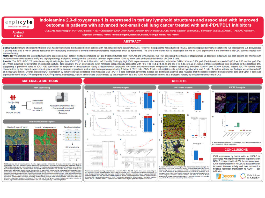
Patients with high IDO1 expression showed significantly improved progression-free survival compared with patients with low IDO1 expression.
High IDO1 expression was also associated with a better objective response rate, with 19.9% in IDO1-high patients compared with 5.2% in IDO1-low patients.
Overall survival was also improved in IDO1-high patients, with a median overall survival of 15.9 months compared with 9.65 months in IDO1-low patients.
After adjustment for covariates, including histological subtype, TLS signature, and PD-L1 expression, IDO1 remained independently associated with improved progression-free survival and overall survival.
These associations were not observed in the docetaxel arm, suggesting that IDO1 gene expression may have predictive value specifically for response to atezolizumab.
Deconvolution analysis showed that the tumor microenvironment composition differed significantly between IDO1-high and IDO1-low tumors.
IDO1-high tumors were characterized by higher expression of genes associated with immune cell populations, including interferon-gamma-producing Th1 cells, CD8+ T cells, natural killer cells, cytotoxic lymphocytes, and B cells.
Multiplex immunofluorescence analysis performed on 53 NSCLC tumors confirmed that elevated IDO1 expression by tumor cells was correlated with increased infiltration of CD8+/PD1+ T cells.
Spatial analysis further showed that the relative distance between tumor cells and CD8+ T cells was significantly lower in IDO1-high tumors compared with IDO1-low tumors.
Interestingly, 52% of tumors were characterized by the presence of tertiary lymphoid structures. IDO1 expression was observed in most TLS analyzed, notably in follicular dendritic cells.
Conclusion
These findings suggest that high IDO1 expression is associated with improved clinical outcome in NSCLC patients treated with atezolizumab.
Rather than acting only as an immunosuppressive marker, IDO1 expression in this context may reflect a pre-existing inflamed tumor microenvironment enriched in cytotoxic immune populations, CD8+/PD1+ T cells, B cells, and tertiary lymphoid structures.
This study highlights the importance of combining transcriptomic analysis, multiplex immunofluorescence, and spatial digital pathology to better understand biomarkers of response to immune checkpoint blockade in NSCLC.