
Background
The tumor microenvironment of epithelial ovarian cancer is unique among solid tumors, as tumor cells can shape their own microenvironment through the formation of malignant ascites.
These ascites represent a complex mixture of soluble factors and cellular components. They are known to be enriched in macrophages, which have been suggested to acquire an M2-like phenotype involved in tumor progression, resistance, and metastasis.
However, tumor-associated macrophages from ovarian cancer patient ascites may also display mixed M1/M2 properties, contributing to a pro-inflammatory and tumor-promoting microenvironment.
Characterizing both the cellular composition and the soluble factors present in ovarian cancer ascites is therefore crucial to better understand ovarian cancer biology and the immunosuppressive pathways that may alter immune cell activity.
Methods
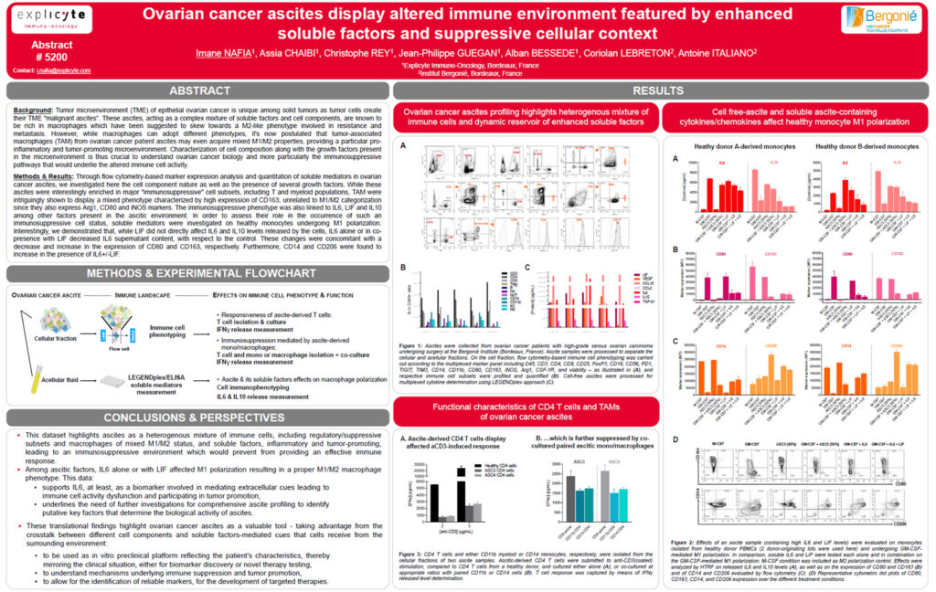
Ovarian cancer ascites were analyzed using flow cytometry-based marker expression profiling to characterize the cellular composition of the ascitic microenvironment.
In parallel, soluble mediators and growth factors present in the ascites were quantified to assess their potential contribution to immune cell polarization and immunosuppressive activity.
To further investigate the role of selected soluble mediators, healthy monocytes undergoing M1 polarization were exposed to factors identified in the ascitic environment, including IL-6 and LIF.
Results
Ovarian cancer ascites were enriched in major immunosuppressive cell subsets, including T cell and myeloid populations.
Tumor-associated macrophages displayed a mixed phenotype characterized by high expression of CD163, together with markers not strictly restricted to classical M1 or M2 categorization, including Arg1, CD80, and iNOS.
This immunosuppressive phenotype was associated with the presence of soluble mediators in the ascitic environment, including IL-6, LIF, and IL-10.
In monocyte polarization experiments, LIF alone did not directly affect IL-6 or IL-10 levels released by the cells.
In contrast, IL-6, either alone or in combination with LIF, decreased IL-6 levels in the supernatant compared with control conditions.
These changes were associated with decreased expression of CD80 and increased expression of CD163, suggesting a shift toward a more immunosuppressive phenotype.
In addition, CD14 and CD206 expression increased in the presence of IL-6, with or without LIF, further supporting the role of ascites-associated soluble mediators in shaping myeloid cell phenotype.
Conclusion
These findings highlight the complex immunosuppressive nature of ovarian cancer ascites, characterized by the presence of regulatory immune cell populations, tumor-associated macrophages with mixed M1/M2 features, and soluble mediators such as IL-6, LIF, and IL-10.
The data suggest that ascites-derived factors, particularly IL-6, may contribute to the acquisition of an immunosuppressive myeloid phenotype.
This work supports the relevance of integrated immune profiling and soluble factor analysis to better understand ovarian cancer biology and identify pathways that may be targeted to restore effective anti-tumor immunity.