
Background
Ovarian cancer ascites are increasingly recognized as important contributors to ovarian cancer progression. The behavior of tumor cells is strongly influenced by the composition of their surrounding microenvironment.
Characterizing the cellular and soluble composition of ascites is therefore essential to better understand how this specific milieu affects tumor progression.
In particular, ascites may contribute to immunosuppressive pathways that alter immune responses and promote immune dysfunction within the tumor microenvironment.
Methods
Ascites samples were collected from patients with advanced ovarian cancer.
The immune context of these samples was profiled using multiplexed approaches, including flow cytometry-based marker expression analysis and quantification of soluble mediators.
Immune cell composition was investigated together with the levels of multiple soluble factors, including cytokines, chemokines, and mediators associated with amino acid metabolic pathways known to be involved in immunosuppression.
In addition, ascites fluids were functionally screened to evaluate their biological effects on healthy monocytes, either in an undifferentiated state or during M1 polarization.
Results
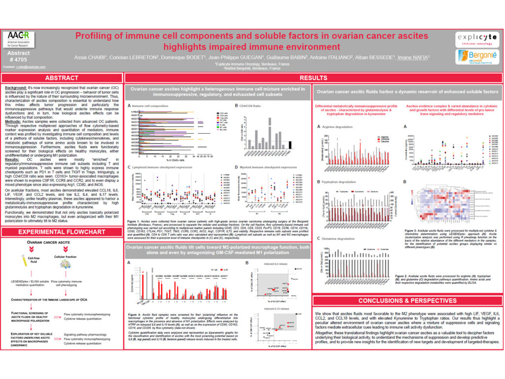
Ovarian cancer ascites were enriched in regulatory and immunosuppressive immune cell subsets, including both T cell and myeloid populations.
T cells showed high expression of immune checkpoints, including PD1 on T cells and TIGIT on regulatory T cells.
A high CD4/CD8 ratio was also observed, further supporting an altered immune landscape within the ascitic microenvironment.
CD163+ tumor-associated macrophages expressed CSF1R, CCR8, and CCR2.
These macrophages also displayed a mixed phenotype, with expression of Arg1, CD80, and iNOS, suggesting that they do not fit strictly into a classical M1 or M2 polarization profile.
Analysis of acellular ascites fractions showed elevated levels of CCL18, IL6, LIF, VEGF, and CCL2 in most samples.
In contrast, IL2, IL4, and IL17 levels were low.
Compared with healthy plasma, ovarian cancer ascites displayed a metabolically immunosuppressive profile characterized by high glutaminolysis and increased tryptophan degradation into kynurenine.
Functional assays demonstrated that ascites were able to polarize monocytes toward an M2-like macrophage phenotype.
Ascites also antagonized M1 polarization, ultimately shifting monocytes toward an M2-like immunosuppressive status.
Conclusion
These findings highlight the immunosuppressive nature of ovarian cancer ascites, characterized by regulatory immune cell populations, immune checkpoint expression, suppressive myeloid features, and metabolic alterations.
The data suggest that ovarian cancer ascites actively shape monocyte and macrophage polarization, promoting an M2-like phenotype and limiting inflammatory M1 differentiation.
This work supports the relevance of integrated immune, soluble mediator, and metabolic profiling to better understand ovarian cancer progression and identify pathways that may be targeted to restore effective anti-tumor immunity.