
Background
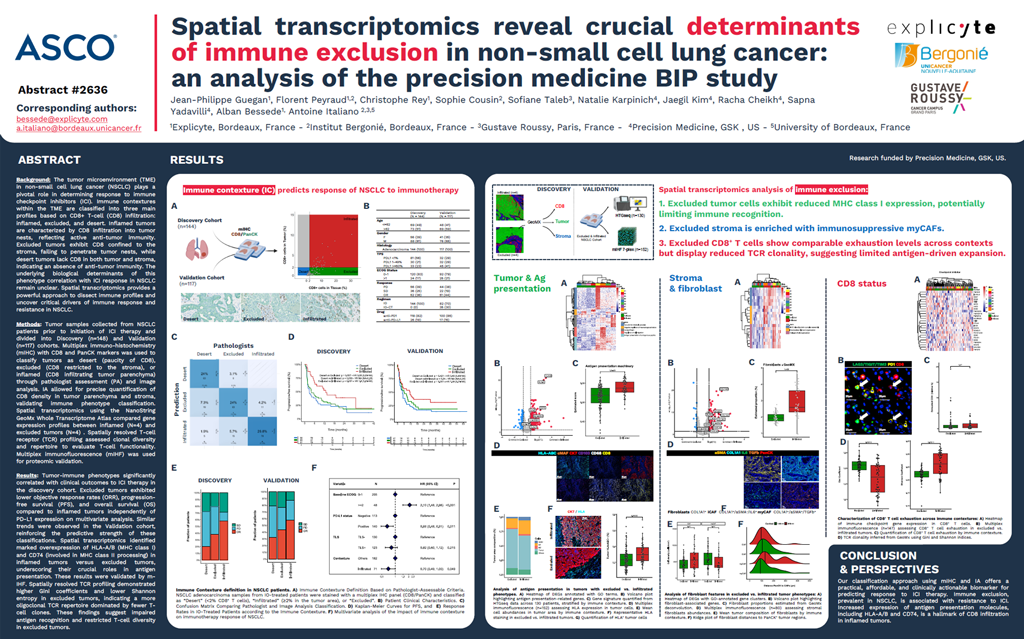
The tumor microenvironment (TME) in non-small cell lung cancer (NSCLC) plays a pivotal role in determining response to immune checkpoint inhibitors (ICI). Immune contextures within the TME are classified into three main profiles based on CD8+ T-cell (CD8) infiltration: inflamed, excluded, and desert. Inflamed tumors are characterized by CD8 infiltration into tumor nests, reflecting active anti-tumor immunity. Excluded tumors exhibit CD8 confined to the stroma, failing to penetrate tumor nests, while desert tumors lack CD8 in both tumor and stroma, indicating an absence of anti-tumor immunity. The underlying biological determinants of this phenotype correlation with ICI response in NSCLC remain unclear. Spatial transcriptomics provides a powerful approach to dissect immune profiles and uncover critical drivers of immune response and resistance in NSCLC.
Methods
Tumor samples collected from NSCLC patients prior to initiation of ICI therapy and divided into Discovery (n=148) and Validation (n=117) cohorts. Multiplex immuno-histochemistry (mIHC) with CD8 and PanCK markers was used to classify tumors as desert (paucity of CD8), excluded (CD8 restricted to the stroma), or inflamed (CD8 infiltrating tumor parenchyma) through pathologist assessment (PA) and image analysis. IA allowed for precise quantification of CD8 density in tumor parenchyma and stroma, validating immune phenotype classification. Spatial transcriptomics using the NanoString GeoMx Whole Transcriptome Atlas compared gene expression profiles between inflamed (N=4) and excluded tumors (N=4). Spatially resolved T-cell receptor (TCR) profiling assessed clonal diversity and repertoire to evaluate T-cell functionality. Multiplex immunofluorescence (mIHF) was used for proteomic validation.
Results
Tumor-immune phenotypes significantly correlated with clinical outcomes to ICI therapy in the discovery cohort. Excluded tumors exhibited lower objective response rates (ORR), progression-free survival (PFS), and overall survival (OS) compared to inflamed tumors independently of PD-L1 expression on multivariate analysis. Similar trends were observed in the Validation cohort, reinforcing the predictive strength of these classifications. Spatial transcriptomics identified marked overexpression of HLA-A/B (MHC class I) and CD74 (involved in MHC class II processing) in inflamed tumors versus excluded tumors, underscoring their crucial roles in antigen presentation. These results were validated by mIHF. Spatially resolved TCR profiling demonstrated higher Gini coefficients and lower Shannon entropy in excluded tumors, indicating a more oligoclonal TCR repertoire dominated by fewer T-cell clones. These findings suggest impaired antigen recognition and restricted T-cell diversity in excluded tumors.