
Background & rationale
- NY-ESO-1 is an immunogenic cancer-testis antigen and a therapeutic target for TCR-based strategies.
- Its value as a baseline biomarker for PD-1/PD-L1 blockade in NSCLC remains poorly defined.
- Because NY-ESO-1 is biologically relevant only when expressed by tumor epithelial cells, this study focused on CK7+/NY-ESO-1+ tumor cells.
Study design
- Cohort 190 baseline FFPE tumors from advanced NSCLC patients treated with PD-1/PD-L1 inhibitors
- Multiplex IF Panel included CD8, CD68, CD103, CK7, HLA-ABC, cMAF, NY-ESO-1 and pSMAD3
- Quantification PhenoImager HT imaging, QuPath segmentation and FlowJo-based phenotyping after signal normalization.
- Clinical endpoints RECIST response, durable clinical benefit, PFS, OS and exploratory biologic analyses.
Exploratory HTG whole-transcriptome analysis used a selected subset.
Clinical plots use cutpoint-defined High vs Low groups based on evaluable CK7+/NY-ESO-1+ tumor burden.
Main clinical findings
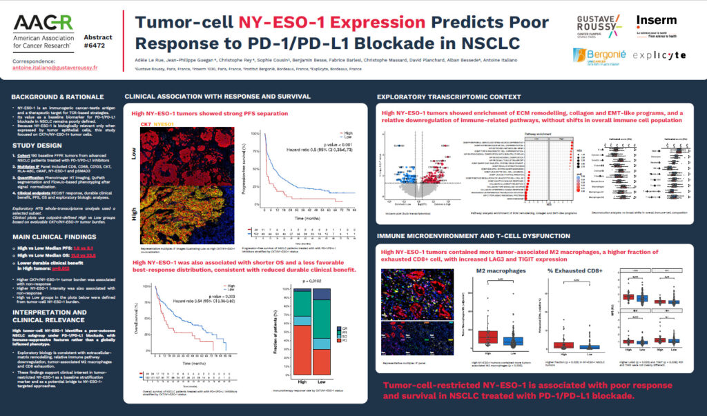
- High vs Low Median PFS: 1.8 vs 8.1
- High vs Low Median OS: 11.0 vs 33.5
- Lower durable clinical benefit in High tumors: p=0.013
- Higher CK7+/NY-ESO-1+ tumor burden was associated with non-response
- Higher NY-ESO-1 intensity was also associated with non-response
- High vs Low groups in the plots below were defined from tumor-cell NY-ESO-1 burden.
Interpretation and clinical relevance
High tumor-cell NY-ESO-1 identifies a poor-outcome NSCLC subgroup under PD-1/PD-L1 blockade, with immune-suppressive features rather than a globally inflamed phenotype.
- Exploratory biology is consistent with extracellular-matrix remodelling, relative immune pathway downregulation, tumor-associated M2 macrophages and CD8 exhaustion.
- These findings support clinical interest in tumor-restricted NY-ESO-1 as a baseline stratification marker and as a potential bridge to NY-ESO-1-targeted approaches.