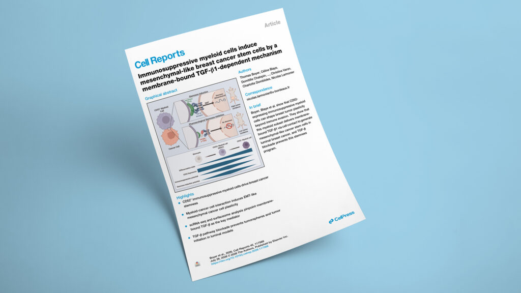
Publication in Cell Reports

How a CD52⁺ myeloid subset drives mesenchymal breast cancer stemness through membrane-bound TGF-β1

Explicyte collaborated with: CHU Bordeaux·CHU de Dijon·CNRS·Inserm·Institut Bergonié·University Marie et Louis Pasteur·University of Bordeaux·University of Montreal
Immunosuppressive myeloid cells induce mesenchymal-like breast cancer stem cells by a membrane-bound TGF-β1-dependent mechanism
BreastDiscoveryTarget discoveryTarget validationFFPE tissueImmune/tumor cell co-culturePBMCscRNAseq
JournalCell Reports
DateJul 2026
Read full paper →

Immunosuppressive myeloid cells are known for shielding tumors from immune attack — but whether they also make cancer cells harder to eradicate has been unclear. Combining single-cell transcriptomics, cell-surface proteomics, and in vivo tumor-initiation assays, the team shows that a CD52-expressing myeloid subset reprograms luminal breast cancer cells into mesenchymal cancer stem cells. The trigger is membrane-bound TGF-β1 delivered through direct cell-to-cell contact — not a secreted factor — pointing to a spatially restricted target that today's soluble-TGF-β inhibitors may miss.

A new study in Cell Reports — led by Nicolas Larmonier (University of Bordeaux, CNRS, INSERM, ImmunoConcEpT UMR5164), with Thomas Boyer and Céline Blaye as co-first authors and Christine Varon and Charlotte Domblides as co-senior authors — shows that immunosuppressive myeloid cells do more than evade immunity: they actively reprogram breast cancer cells toward a stem-like, mesenchymal state. The work drew on malignant pleural effusions collected from breast cancer patients at Institut Bergonié and was supported by the French National League Against Cancer, SIRIC-BRIO, the ARC Foundation, ITMO Aviesan (ANR), and the GILEAD Research Scholars Program.

The question

Can immunosuppressive myeloid cells turn breast cancer cells into mesenchymal cancer stem cells — and what molecular signal drives it?

Key steps

  1. 1

    Myeloid cells expand breast cancer stem cells

    Human monocyte-derived suppressor cells (HuMoSC) increased MCF7 tumorsphere formation in a ratio-dependent way, while non-suppressive monocytes did not. Extreme limiting dilution assays put the stem-cell frequency at 1/25.1 with HuMoSC versus 1/45.5 for cancer cells alone (P < 0.00187), and flow cytometry showed rises in both CD44⁺CD24⁻ and ALDH⁺ populations. In orthotopic NSG xenografts, co-culture raised the tumor-initiating cell frequency to 1/962 versus 1/2,313 for MCF7 alone. The effect held in luminal lines (MCF7, T47D) but was absent in triple-negative lines (BT549, MDA-MB-231).

  2. 2

    Single-cell RNA-seq resolves a mesenchymal stem-cell state

    scRNA-seq of 15,902 cells (8,643 HuMoSC and 7,259 MCF7) across 2D, tumorsphere, and co-culture conditions yielded four MCF7 clusters. Co-culture selectively enriched a mesenchymal cancer-stem-cell cluster marked by high ZEB1, VIM, POU5F1/OCT4, and NOTCH1 with low CD24 (M_CSC), rather than the epithelial CSC cluster — indicating myeloid cells push plasticity toward the invasive mesenchymal state.

  3. 3

    A CD52⁺ subset does the work

    Differential expression flagged CD52 as the surface marker separating two core HuMoSC subsets, confirmed by flow cytometry. Purified CD52⁺ HuMoSC were immunosuppressive and induced tumorspheres, while CD52⁻ cells lacked both properties. In patient pleural effusions (eight collected 2022–2025; seven usable), only CD33^high CD52⁺ cells promoted stemness, extending the finding to primary cells.

  4. 4

    Transcriptome and surfaceome converge on TGF-β1

    CellChat ligand-receptor inference across the eight clusters, combined with a biotinylation-based surface proteome (611 surface proteins on HuMoSC, 488 on MCF7 by LC-MS/MS), narrowed the candidates to TGFβ and MHC-II. Surfaceome data showed high surface TGF-β1 on HuMoSC and its receptors TGF-βR1 and ACVR1 on MCF7; the TGFβ pathway was also enriched in bulk RNA-seq, giving orthogonal support.

  5. 5

    Membrane-bound TGF-β1 is necessary and sufficient

    Transwell separation and conditioned medium both failed to induce stemness, showing contact is required. PFA-fixed HuMoSC still induced tumorspheres — and pre-treating them with an anti-TGF-β antibody abolished it — demonstrating the membrane-bound ligand is sufficient. The TGF-βRI/II inhibitor Ly2109761 and a pan-TGF-β capture antibody blocked induction by both HuMoSC and patient-derived CD33^high CD52⁺ cells.

Impact

The findings recast immunosuppressive myeloid cells as active drivers of cancer stemness rather than passive immune shields, and point to a contact-dependent, membrane-tethered form of TGF-β1 that behaves differently from the soluble cytokine most therapies target.

1/962
tumor-initiating cell frequency after myeloid co-culture, vs 1/2,313 for cancer cells alone (in vivo)
CD52⁺
the single myeloid subset that induces stemness — CD52⁻ counterparts had no effect
contact-only
stemness required direct cell contact; conditioned medium and transwell co-culture were inactive

For drug developers, this positions membrane-bound TGF-β1 as a spatially restricted target that soluble-TGF-β inhibitors may fail to engage — arguing for agents that reach the ligand at the myeloid–tumor interface. The strict luminal-subtype dependence is a stratification signal: TNBC models with pre-existing mesenchymal programs did not respond. CD52 offers a candidate handle on the responsible myeloid population, though the authors caution it is context-dependent and insufficient on its own to define a homogeneous subset.

Mapping myeloid–tumor crosstalk or a membrane-bound TGF-β program in your breast cancer models? Let's talk.

Talk to us
Explicyte Oncology CRO logo

Capabilities

Modalities