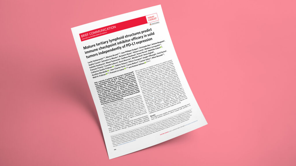
Publication in Nature Cancer

How mature TLSs identify solid tumor patients more likely to benefit from PD-1 blockade

Mature tertiary lymphoid structures predict immune checkpoint inhibitor efficacy in solid tumors independently of PD-L1
JournalNature Cancer
DateAug 2021
Read full paper →

Checkpoint inhibitors remain hard to predict: PD-L1 misses many responders and nonresponders. In an AstraZeneca-funded, Bordeaux-led Nature Cancer analysis, Explicyte-affiliated authors helped evaluate mature tertiary lymphoid structures across 540 pre-treatment FFPE tumors from three anti-PD-1/PD-L1 cohorts. Mature TLSs correlated with higher response, longer PFS and longer OS independently of PD-L1 and CD8 density, supporting TLS maturity as a practical tissue biomarker for patient stratification.

Explicyte-affiliated authors Jean-Philippe Guégan, Christophe Rey, Félicie Courgeon, and Alban Bessede contributed to this Nature Cancer paper, with Alban Bessede co-conceiving and co-designing the analysis and contributing to the TLS immunology assessment, figure and table development, literature synthesis, and manuscript writing. The work was led by Institut Bergonié and the University of Bordeaux, with major collaborators including Centre de Recherche des Cordeliers, Gustave Roussy, Clinique Marzet, and AstraZeneca. The study was funded by AstraZeneca and built around three independent anti-PD-1/PD-L1-treated solid tumor cohorts, including the Institut Bergonié institutional tumor-profiling program NCT02534649 and the MATCH-R program NCT02517892. The central question was translational: whether a tissue feature visible in routine pathology and multiplex immunophenotyping could identify patients likely to benefit from immune checkpoint blockade beyond PD-L1 status alone.

The question

Can mature tertiary lymphoid structures predict checkpoint inhibitor benefit in solid tumors independently of PD-L1 and CD8 density?

Key steps

  1. 1

    Assemble three ICI cohorts

    The analysis used pre-treatment FFPE tumor material from 328 patients in the discovery cohort, 131 patients in validation cohort A, and 81 patients in validation cohort B, for 540 tumors overall. All patients had advanced solid tumors treated with anti-PD-1 or anti-PD-L1 antibodies and at least one post-treatment imaging evaluation. The cohorts covered multiple histologies, including NSCLC, soft-tissue sarcoma, bladder, colorectal, head and neck, renal, breast, thyroid, pancreatic, gynecologic, and gastric cancers.

  2. 2

    Score TLS maturity in tissue

    TLS status was assessed on serial FFPE sections using HES, CD3/CD20 double IHC, CD8/PD-L1 double IHC, and multiplex immunofluorescence. TLS maturity was evaluated with a CD4, CD8, CD20, CD21, and CD23 panel, with mature TLSs defined by CD23+ follicular dendritic cells. Staining and imaging workflows used Ventana Discovery Ultra, Ventana BenchMark ULTRA, Akoya Opal fluorophores, and Vectra Polaris scanning, with CD8+ T-cell density quantified digitally.

  3. 3

    Link mTLSs to outcomes

    In the 328-patient discovery cohort, TLSs were detected in 105 tumors (32%), including 84 mature TLS-positive tumors (25.6%). Objective response occurred in 31 of 84 patients with mTLS-positive tumors (36.9%), compared with 4 of 21 patients with immature TLSs (19.3%) and 43 of 223 patients with no TLSs (19.0%; P = 0.015). Median PFS was 6.1 months in the mTLS-positive group versus 2.7 months in the mTLS-negative group (P = 0.015), and median OS was 24.8 versus 13.3 months (P = 0.016).

  4. 4

    Test independence from PD-L1

    PD-L1 TPS was ≥1% in 70 of 328 discovery-cohort tumors (21.3%), with similar PD-L1 positivity across mTLS-high and mTLS-low or mTLS-absent tumors. In PD-L1-negative tumors, objective response reached 35.6% with mTLSs versus 14.1% without mTLSs; in PD-L1-positive tumors, response reached 69.2% versus 40.4%. In multivariate analyses adjusted for clinical and immune variables, mTLS status retained independent predictive value for PFS and OS.

  5. 5

    Validate clinical robustness

    Validation cohort A confirmed higher objective response in mTLS-positive tumors than mTLS-negative tumors: 50.0% versus 27.6% (P = 0.009), with median PFS of 8.0 versus 3.5 months (P = 0.038). Validation cohort B showed the same direction of effect, with objective response of 38.4% versus 11.8% (P = 0.02) and median OS of 24.6 versus 8.1 months (P = 0.036). A routine-compatible CD20/CD23 double IHC approach matched multiplex immunofluorescence results in 91.9% of assessed cases.

Impact

Mature TLSs add a structured B-cell and follicular dendritic-cell dimension to checkpoint inhibitor biomarker analysis. The findings support TLS maturity as a tissue-based readout that can complement PD-L1, CD8 density, and other tumor-intrinsic biomarkers.

540
pre-treatment FFPE tumors analyzed across one discovery cohort and two independent validation cohorts
36.9%
objective response rate in mTLS-positive tumors in the discovery cohort, versus 19.0% in tumors with no TLSs
91.9%
concordance between CD20/CD23 double IHC and multiplex immunofluorescence for TLS maturity assessment

For clinical biomarker teams, mature TLSs offer a practical way to evaluate whether a tumor microenvironment is organized for response to PD-1/PD-L1 blockade, not simply infiltrated by immune cells. For drug developers, TLS maturity can help enrich trial populations, interpret discordant PD-L1 results, and design combination strategies that aim to generate or sustain B-cell-rich immune niches in tumors.

Designing an ICI biomarker study around TLS maturation and B-cell niches? Let’s talk.

Talk to us
Explicyte Oncology CRO logo

Capabilities

Modalities