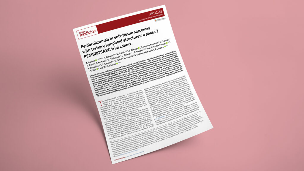
Publication in Nature Medicine

How tertiary lymphoid structures predict pembrolizumab response in soft-tissue sarcoma — and why plasma cells matter

Pembrolizumab in soft-tissue sarcomas with tertiary lymphoid structures: a phase 2 PEMBROSARC trial cohort
JournalNature Medicine
DateMay 2022
Read full paper →

Checkpoint inhibitors barely move the needle in unselected advanced soft-tissue sarcoma — objective responses hover near 2%. The TLS-positive cohort of the phase 2 PEMBROSARC trial tested a different approach: enrolling only patients whose tumors already contained tertiary lymphoid structures. In these 30 patients, pembrolizumab plus low-dose cyclophosphamide produced a 30% objective response rate and met its primary endpoint. Spatial transcriptomics and multiplex imaging then traced response to intratumoral plasma cells and dendritic cells — pointing toward a biomarker-driven selection strategy for sarcoma immunotherapy.

Published in Nature Medicine, this PEMBROSARC trial cohort was led by Prof. Antoine Italiano (Institut Bergonié, University of Bordeaux) — with Alban Bessede of Explicyte as a co–first author — and drew on the French Sarcoma Group together with the tertiary-lymphoid-structure biology group of Catherine Sautès-Fridman and Wolf Hervé Fridman at the Centre de Recherche des Cordeliers. The work was supported by the RHU CONDOR program and by MSD, France’s Agence Nationale de la Recherche, INSERM, the Ligue contre le Cancer, and the French National Cancer Institute. Explicyte contributed the exploratory biomarker workup that explains who responds: spatial transcriptomic profiling on the NanoString GeoMx whole-transcriptome atlas and multiplex immunofluorescence panels for TLS imaged on the PhenoImager HT, applied to responder and non-responder tumors.

The question

Can the presence of tertiary lymphoid structures identify the soft-tissue sarcoma patients most likely to benefit from pembrolizumab — and which immune cells explain who responds?

Key steps

  1. 1

    Select patients by tertiary lymphoid structures

    Of 240 advanced soft-tissue sarcomas screened for TLS status, 48 (20%) were TLS-positive, defined as a CD3+/CD20+ aggregate of at least 700 cells in the tumor bed. Thirty TLS-positive patients were assessable for efficacy after treatment with pembrolizumab plus low-dose cyclophosphamide. TLS-positive cases were spread across histologies, with higher rates in dedifferentiated liposarcoma than in leiomyosarcoma.

  2. 2

    Primary endpoint met, responses ~10× the unselected rate

    The 6-month non-progression rate was 40% (95% CI, 22.7–59.4), meeting the primary endpoint, and the objective response rate reached 30% (95% CI, 14.7–49.4). In the trial’s earlier all-comer cohorts the same regimen produced a 4.9% non-progression rate and a 2.4% response rate — and all but one of those patients were later found to be TLS-negative. Median OS in the TLS-positive cohort was 18.3 months and median duration of response was 11.0 months.

  3. 3

    Spatial profiling separates responders from non-responders

    Explicyte ran spatial transcriptomics on the NanoString GeoMx whole-transcriptome atlas (>18,000 protein-coding genes) across six tumors — three responders, three with progressive disease — segmenting regions into B-cell (CD20+) versus other-immune areas inside TLSs and tumor-versus-stroma (CD45+) areas. Deconvolution (SpatialDecon) showed responder stroma significantly enriched in plasma cells, while TLSs of non-responders were enriched in regulatory T cells.

  4. 4

    Multiplex imaging pinpoints the cells that matter

    Three multiplex immunofluorescence panels (CD8/GzmA/CD4/FOXP3/CD56; MUM1/CD20/CD27/CD3/COL1A1/IgG; HLA-DR/CD11c/CD1c/CD68/CD83), stained on the Ventana Discovery platform with Akoya Opal reagents and imaged on the PhenoImager HT, profiled whole tumor sections. IgG-expressing MUM1+ plasma cells marked responders — tumors highly infiltrated by plasma cells had a median PFS of 13.8 months versus 2.6 months in low-infiltration cases (P = 0.009). High CD11c+/HLA-DR+ dendritic-cell density tracked with better PFS and OS, while CD4+/FOXP3+ Treg-rich TLSs marked poorer outcome.

Impact

Selecting sarcoma patients by TLS status converts a treatment with negligible activity into one with meaningful, durable responses — and the immune profiling nominates plasma cells as a refinement beyond TLS status alone.

30%
objective response rate in TLS-positive sarcomas (vs 2.4% in unselected patients)
20%
of advanced soft-tissue sarcomas are TLS-positive
13.8 mo
median PFS in plasma-cell–high tumors vs 2.6 months when low

For sponsors developing immunotherapy in sarcoma, TLS status offers a prospectively validated enrichment strategy that can rescue trials otherwise undermined by low unselected response rates. The plasma-cell and regulatory-T-cell signals point toward a next-generation biomarker panel and rational combinations — pairing anti–PD-1 with Treg- or CTLA-4–directed agents. Two randomized CONDOR-associated trials are already testing TLS-based selection in the neoadjuvant and metastatic settings.

Building a TLS- or plasma-cell–driven patient-selection strategy for a sarcoma immunotherapy trial? Let's talk.

Talk to us
Explicyte Oncology CRO logo

Capabilities

Modalities