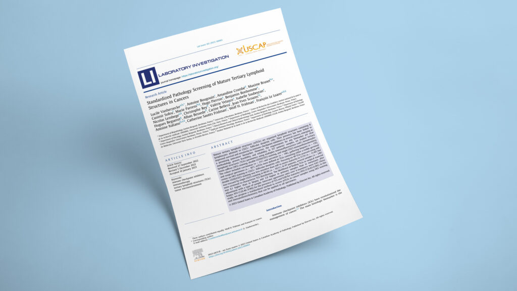
Publication in Laboratory Investigation

How to screen for mature tertiary lymphoid structures in routine pathology — a standardized method validated across 357 tumors

Standardized Pathology Screening of Mature Tertiary Lymphoid Structures in Cancers
JournalLaboratory Investigation
DateJan 2023
Read full paper →

Mature tertiary lymphoid structures (mTLS) — B-cell follicles with CD23+ follicular dendritic cells — predict response to immune checkpoint inhibitors across many cancers, sometimes outperforming PD-L1 and tumor mutational burden. But their promise depends on being able to score them reliably in an ordinary pathology lab, and criteria have varied study to study. Across 357 carcinoma and sarcoma samples, this study built and validated a routine-technique workflow — HES staining plus CD20 and CD23 immunohistochemistry — that detects mTLS as sensitively as the multiplex immunofluorescence used in research, with near-perfect agreement between pathologists on maturity. The result is a portable, standardized method ready for clinical trial use.

This validation study in Laboratory Investigation, led by Dr. Lucile Vanhersecke and Dr. François Le Loarer at Institut Bergonié with the Cordeliers immunology group of Prof. Wolf H. Fridman and Prof. Catherine Sautès-Fridman and Prof. Antoine Italiano, set out to make mature tertiary lymphoid structure (mTLS) scoring reproducible in routine pathology. Building on the team’s earlier retrospective and prospective PEMBROSARC-linked work showing mTLS predict checkpoint-inhibitor benefit, it benchmarks a conventional-stain workflow against the multiplex immunofluorescence used in the original research setting. Explicyte performed the blinded multiplex immunofluorescence analysis that served as the reference standard against which the routine-pathology method was validated.

The question

Can mature tertiary lymphoid structures be scored as reliably with routine pathology stains as with research-grade multiplex immunofluorescence — reproducibly enough to use as a clinical biomarker?

Key steps

  1. 1

    Set the multiplex-IF reference standard

    The routine-pathology workflow was validated against multiplex immunofluorescence as the benchmark. Explicyte analyzed the multiplex-IF assays blind (with W.H. Fridman), scoring CD20/CD21/CD23 whole-slide images in HALO software to identify follicular dendritic cell networks and call TLS maturity. This mIF read-out was the reference against which HES and CD20/CD23 immunohistochemistry were measured across the cohort.

  2. 2

    Conventional stains matched multiplex IF for maturity

    In 62 TLS-positive samples scored blind by both methods, all 30 mTLS called by IHC were confirmed by multiplex IF, and 40 of 42 (95.2%) mIF-mature cases were confirmed by IHC. Where double CD20/CD23 staining missed follicular dendritic cells (11 of 40 cases, 27.5%), adding a single CD23 stain rescued the maturity call in 10 of 11 (90.9%) — establishing single CD20 plus single CD23 as the most sensitive routine combination, with HES-visible germinal centers needing no immunostaining at all.

  3. 3

    Sampling drives whether TLS are found

    Reviewing 240 samples from 97 TLS-positive patients, the team showed detection depends heavily on the specimen. Surgical resections were 6.1× more likely to contain TLS than biopsies (95% CI 3.5–10.7; P < 0.0001), and primary tumors about 2× more likely than metastases. TLS also clustered at the invasive tumor front rather than the tumor core — practical guidance for where and what to sample.

  4. 4

    The method is reproducible across pathologists

    Four pathologists (two trainees, two seniors) scored a 67-sample set after brief training. Interrater agreement was substantial for TLS presence (Fleiss κ 0.65) and near-perfect for maturity (Fleiss κ 0.90). Residual disagreements centered on interpreting isolated CD23+ cells, prompting the authors to publish a shared “atlas of TLS” to standardize these gray-zone calls.

Impact

The study closes the gap between a research biomarker and a clinical one: mTLS can now be scored the same way, sensitively and reproducibly, in any pathology lab with standard stains.

357
tumor samples (carcinomas and sarcomas) used to build and validate the screening method
κ 0.90
interrater agreement on TLS maturity across four pathologists (near-perfect)
91%
of double-stain–missed cases rescued by adding a single CD23 stain

For trial sponsors and pathology labs, this provides a validated, low-cost path to use mTLS status as a patient-selection biomarker without specialized multiplex platforms — increasingly relevant as mTLS enters immunotherapy trials as an inclusion criterion (e.g., NCT04095208, NCT04874311, NCT04968106). The sampling findings matter operationally: biopsies and metastatic samples under-detect TLS, so trial design and reporting should standardize specimen type, and scoring should default to IHC when crush artifact degrades HES. A shared training atlas is the mechanism for keeping scoring consistent across sites.

Need reproducible ISO-certified mature-TLS scoring — multiplex IF or a routine-IHC workflow — to stratify patients for an immunotherapy trial? Let's talk.

Talk to us
Explicyte Oncology CRO logo

Capabilities

Modalities