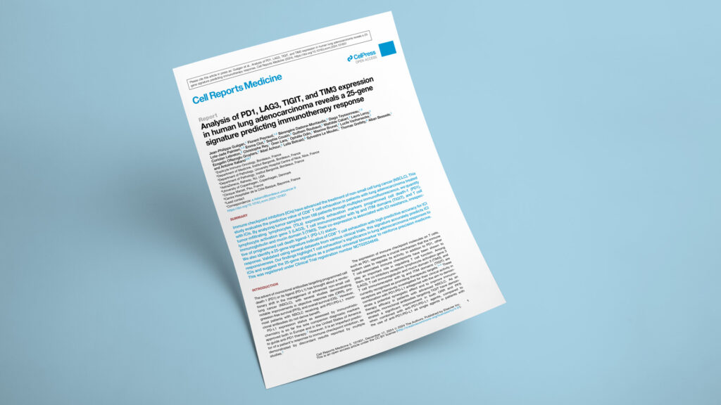
Publication in Cell Reports Medicine

Why CD8 T-cell exhaustion predicts immunotherapy resistance in lung cancer — and a 25-gene signature that flags it

Explicyte collaborated with: AstraZeneca·Centre Hospitalier de la Côte Basque·CHU Nice·Clinique Marzet·Institut Bergonié·University of Copenhagen
Analysis of PD1, LAG3, TIGIT, and TIM3 expression in human lung adenocarcinoma reveals a 25-gene signature predicting immunotherapy response
GenitourinarySkinThoracicBiomarker analysisBiomarker discoveryDiscoveryTrialsFFPE tissueBioinformaticsMultiplex IF/IHCRNAseq
JournalCell Reports Medicine
DateDec 2024
Read full paper →

Most patients with advanced lung cancer never respond to PD-1/PD-L1 checkpoint inhibitors, and PD-L1 staining alone doesn't reliably say who will. Profiling tumors from 166 lung adenocarcinoma patients with a six-marker multiplex immunofluorescence panel — then whole-transcriptome sequencing of 135 of them — the team found that CD8+ T cells co-expressing PD1 with LAG3, TIGIT, or TIM3 (an exhausted phenotype) mark tumors that resist immunotherapy, independent of PD-L1 status. From this they built a 25-gene exhaustion signature that predicted checkpoint-inhibitor response in lung cancer and, across external trial datasets, in melanoma and renal cancer too — pointing toward a pan-tumor immunotherapy biomarker.

A study in Cell Reports Medicine — with Jean-Philippe Guégan (Explicyte) as first author and led by Prof. Antoine Italiano (Institut Bergonié) — asks why so many lung adenocarcinoma patients fail to benefit from PD-1/PD-L1 checkpoint inhibitors, and whether the exhaustion state of their CD8+ T cells holds the answer. The work draws on the institutional Bergonié Institute Profiling program (BIP, NCT02534649) and was funded by AstraZeneca and the Nouvelle-Aquitaine Regional Council (Conseil Régional Aquitaine). Explicyte contributed the six-marker multiplex immunofluorescence profiling and the whole-transcriptome (HTG) analysis from which the 25-gene signature was derived.

The question

Does the exhaustion state of CD8+ T cells predict which lung adenocarcinomas will resist immune checkpoint inhibitors — and can it be captured in a portable gene signature?

Key steps

  1. 1

    Multiplex IF maps exhausted CD8 T cells

    Explicyte applied a six-marker multiplex immunofluorescence panel (CD8, CK7, LAG3, PD1, TIGIT, TIM3) using Akoya Opal reagents, Ventana OmniMap detection, and PhenoImager HT imaging to pretreatment tumors from 166 advanced lung adenocarcinoma patients treated with anti-PD1/PD-L1 therapy. Tumors heavily infiltrated by CD8+ cells co-expressing PD1 plus at least one of LAG3, TIGIT, or TIM3 (CD8+/Exh) showed lower response rates (33.3% vs 54.8%; P = 0.01), shorter PFS (median 3 vs 9 months; P = 0.025), and shorter OS (15.4 vs 35.1 months; P = 0.011).

  2. 2

    Exhaustion resists independently of PD-L1

    On multivariate analysis, high CD8+/Exh abundance independently predicted worse PFS (HR 1.66, 95% CI 1.13–2.45; P = 0.010) and OS (HR 1.98, 95% CI 1.31–3.00; P = 0.001), regardless of age, sex, ECOG status, prior treatment lines, or PD-L1 status. PD-L1 positivity, tumor mutational burden, and KRAS and other oncogene mutations were evenly distributed between high- and low-exhaustion tumors — marking exhaustion as a distinct axis of resistance, not a proxy for existing markers.

  3. 3

    A 25-gene signature from HTG transcriptomics

    Explicyte’s translational team profiled 135 FFPE tumors — each already scored for exhaustion by multiplex IF on serial sections — on the HTG transcriptome panel (>19,000 targets; a nuclease-protection assay read out on HTG EdgeSeq). A LASSO model distilled the differentially expressed, PFS-associated genes into a 25-gene exhaustion signature that predicted CD8+ exhaustion with an AUC of 0.98 in the discovery cohort and 0.79 in validation, and separated patients by PFS (P = 0.014).

  4. 4

    Validation across trials and tumor types

    In the POPLAR/OAK atezolizumab arm the signature predicted PFS (HR 0.73; P = 0.003) but did nothing in the docetaxel arm (HR 1.13; P = 0.274), confirming immunotherapy specificity, and it validated in the MATCH-R NSCLC cohort. Beyond lung, a low exhaustion signature predicted better ICI PFS in melanoma (CA209-038; HR 0.30; P = 0.004) and renal cell carcinoma (JAVELIN Renal 101 avelumab-axitinib arm; HR 0.63; P = 0.008), but not in the sunitinib TKI arm — echoing the immunotherapy-specific pattern.

Impact

Immune exhaustion has long been read as a sign of poor prognosis; this work reframes it as a predictive, immunotherapy-specific biomarker — and packages it into a 25-gene test that carries across tumor types.

166
lung adenocarcinoma patients profiled by multiplex IF for T-cell exhaustion
25 genes
exhaustion signature predicting immune checkpoint inhibitor response
AUC 0.98
signature accuracy for CD8+ exhaustion in the discovery cohort

For drug developers, the signature offers a molecularly defined way to enrich immunotherapy trials — and a ready-made rationale for the bispecific antibodies (PD1 × LAG3/TIGIT/TIM3) now in development, whose intended target population is precisely the exhausted-T-cell tumors it identifies. Because it reads out on a scalable bulk-transcriptomic assay rather than single-cell sequencing, it is plausible as a clinical diagnostic. Its performance in melanoma and renal cancer points toward use as a pan-tumor immunotherapy biomarker, pending prospective validation.

Building or validating a T-cell exhaustion or ICI-response signature? Let's talk about pairing multiplex IF with transcriptomic profiling.

Talk to us
Explicyte Oncology CRO logo

Capabilities

Modalities