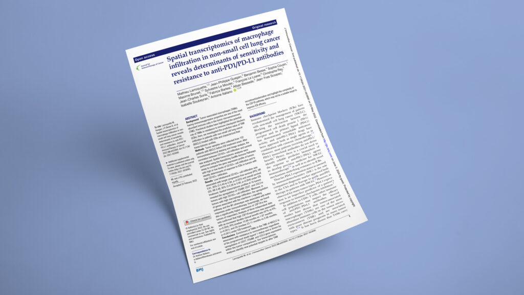
Publication in Journal for ImmunoTherapy of Cancer

Why intratumoral CD163+ macrophages predict anti-PD-1 resistance in non-small cell lung cancer

Spatial transcriptomics of macrophage infiltration in non-small cell lung cancer reveals determinants of sensitivity and resistance to anti-PD1/PD-L1 antibodies
JournalJournal for ImmunoTherapy of Cancer
DateMay 2022
Read full paper →

PD-L1 alone cannot explain why most patients with advanced non-small cell lung cancer fail to benefit from checkpoint blockade. Across 152 pretreatment tumors, the team combined quantitative CD8/CD163 pathology with GeoMx spatial transcriptomics in 16 selected cases. High intratumoral CD163+ macrophage density tracked with shorter survival, while responder tumors with heavy macrophage infiltration retained IFN-γ/M1 programs and unexpectedly higher CSF1R expression. The findings support spatial macrophage profiling while cautioning against blanket CSF1R inhibition.

Published in the Journal for ImmunoTherapy of Cancer, the work was led by Mathieu Larroquette and Jean-Philippe Guégan, who contributed equally, with Antoine Italiano as corresponding author. Explicyte contributed NanoString GeoMx Digital Spatial Profiling to map immune-gene expression separately within PanCK-defined tumor and stromal compartments. This spatial layer helped distinguish pathways associated with macrophage abundance from those linked to checkpoint-blockade benefit within macrophage-rich tumors.

The question

Can the abundance, location, and transcriptional state of tumor-associated macrophages predict response to PD-1/PD-L1 blockade in NSCLC?

Key steps

  1. 1

    Quantify macrophages before immunotherapy

    The team analyzed pretreatment FFPE tumors from 152 patients with advanced NSCLC using CD8, CD163, and CK7 staining on the Ventana Discovery XT platform, followed by PhenoImager HT digitization and InForm image analysis. High intratumoral CD163+ density, defined as more than 472 cells/mm², remained independently associated with shorter progression-free survival (HR 1.80; P = 0.019) and overall survival (HR 2.69; P = 0.002). Stromal CD163+ density did not show the same survival association.

  2. 2

    Map macrophage-associated spatial programs

    Explicyte profiled 16 surgical FFPE tumors on the NanoString GeoMx Immune Pathways assay: eight with low and eight with high CD163+ infiltration. Eleven to 12 regions per case were segmented into PanCK-positive tumor and PanCK-negative stromal compartments, generating 381 spatially distinct areas of illumination. High-macrophage tumor regions showed increased ITGAM, CCL5, and CD27 expression; bulk RNA-seq from 29 cases confirmed their positive correlation with CD163+ density.

  3. 3

    Resolve responder macrophage states

    The team then compared eight macrophage-rich tumors—four from responders and four from patients with progressive disease. Ten stromal genes differed between the groups. Responders showed stronger IFN-γ and M1-associated programs involving STAT1, CD44, IFNGR1, HLA-E, and LY6E, whereas the M2-associated gene BCL2 was higher in nonresponders. CSF1R was unexpectedly among the strongest responder-associated signals.

  4. 4

    Validate the CSF1R association

    In bulk RNA-seq data from 49 patients, high CSF1R expression was associated with a 47% durable clinical benefit rate versus 6% in the low-expression group. Median progression-free survival was 10.89 versus 1.67 months, and median overall survival was 23.11 versus 2.66 months. Two independent immunotherapy-treated NSCLC datasets supported the association, while a 506-patient surgically treated TCGA cohort showed no general prognostic effect.

Impact

Macrophage burden alone does not fully capture immunotherapy biology. Their position relative to tumor cells and their transcriptional state may separate suppressive macrophage-rich tumors from those that remain responsive to checkpoint blockade.

152
Pretreatment NSCLC tumors assessed by quantitative CD8/CD163 pathology
381
Spatial tumor and stromal areas profiled across 16 FFPE tumors by GeoMx DSP VALUE: 47% vs 6%
47% vs 6%
Durable clinical benefit with high versus low CSF1R expression in the RNA-seq cohort

For translational and biomarker teams, the results support measuring where macrophages sit and which immune programs they express—not simply reporting total abundance. For drug developers, the association between higher CSF1R expression and improved checkpoint-blockade outcomes argues against treating CSF1R as a universally suppressive target and supports more phenotype-specific macrophage strategies.

Planning a spatial biomarker program to distinguish macrophage-driven immunotherapy resistance in NSCLC? Let’s talk.

Talk to us
Explicyte Oncology CRO logo

Capabilities

Modalities