Multiplex IF TLS detection & scoring for oncology clinical trials

Explicyte provides validated multiplex IF-based TLS detection and scoring for FFPE tumor samples in oncology studies and clinical trials. Our pathologist-led service delivers standardized TLS classification and study-ready reporting within our ISO 13485 and ISO 9001-certified quality-management framework.

EXPERTS IN TLS EVALUATION

Deploying TLS assays in peer-reviewed publications since 2021  
TLS ASSAY SPECIFICATIONS

A validated TLS assay for oncology clinical trials

Assay
Multiplex immunofluorescence with standardized, pathologist-led TLS interpretation
Intended study use
Evaluation of TLS presence and maturation status in FFPE tumor samples from oncology studies and clinical trials
Reported TLS status
TLS-negative · immature TLS · mature TLS
Multiplex IF panel
CD4 · CD8 · CD20 · CD21 · CD23
Orthogonal morphology / IHC
HES and CD3/CD20 dual IHC on serial sections
Samples
FFPE tumor blocks or unstained slides; tissue adequacy and section requirements confirmed during study setup
Staining & imaging
Automated Ventana Discovery staining · multispectral PhenoImager acquisition
Deliverables
Per-case TLS status report, representative supporting images and cohort-level summary when requested
ISO9001 ISO13485 explicyte biomarker services

Explicyte is ISO 13485:2016 and ISO 9001:2015 certified by Euro-Quality System (certificates 260133/1637F/1 and 260133/1637F/2). The TLS scoring assay is the first biomarker assay developed under this certified QMS, whose scope includes the design and development of tissue biomarkers and tissue-biomarker testing for precision-oncology clinical trials. Assay methods, controls, acceptance criteria, scoring procedures and study records are managed through controlled documentation.

Certification applies to the quality-management system and its defined scope; it does not, by itself, make the TLS assay a CE-marked IVD or companion diagnostic.

How TLS status is classified

Scoring mature versus immature TLS in tumor tissue

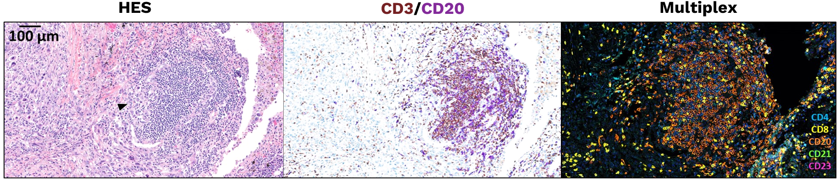
TLS status is assigned by trained pathologists using tissue morphology, lymphoid organization and the CD4/CD8/CD20/CD21/CD23 multiplex IF profile. Mature TLS show germinal-center maturation, identified by a visible germinal center on HES and/or a CD23-positive follicular dendritic-cell network, whereas immature TLS lack these maturation features.
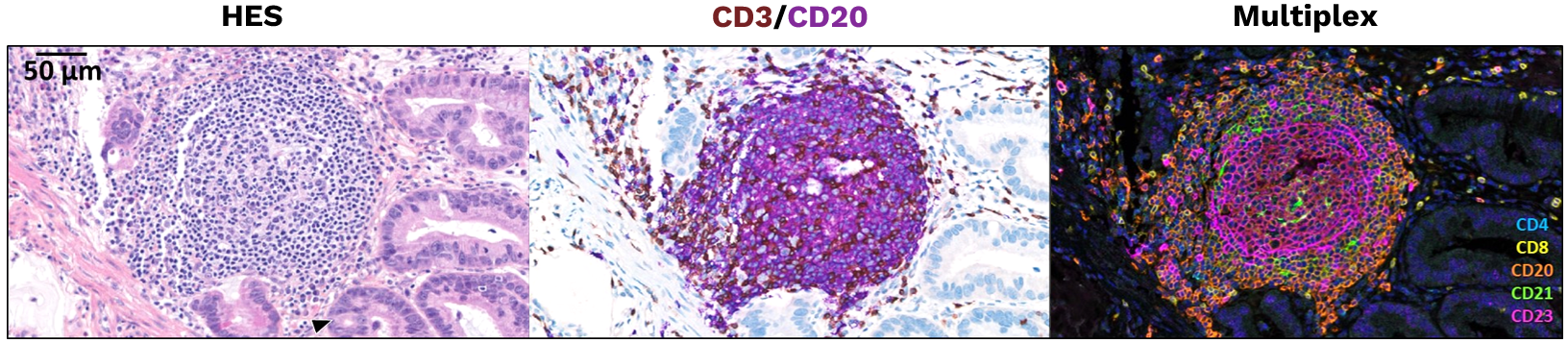
Mature tertiary lymphoid structure in pancreatic adenocarcinoma shown by HES, CD3/CD20 IHC and CD4/CD8/CD20/CD21/CD23 multiplex IF

Mature TLS — primary pancreatic adenocarcinoma

HES, dual IHC (CD3/CD20) and multiplex IF (CD4, CD8, CD20, CD21, CD23). Scale bar: 50 µm.

Immature tertiary lymphoid structure in lung adenocarcinoma shown by HES, CD3/CD20 IHC and CD4/CD8/CD20/CD21/CD23 multiplex IF

Immature TLS — primary lung adenocarcinoma

HES, dual IHC (CD3/CD20) and multiplex IF (CD4, CD8, CD20, CD21, CD23). Scale bar: 100 µm.

TLS ASSAY WORKFLOW

From FFPE tumor tissue to a pathologist-reviewed TLS result

A controlled pathology workflow from sample receipt through staining, imaging, TLS classification and study reporting.

Sample intake & pathology QC

Confirm tissue suitability and prepare serial sections

FFPE tumor blocks or unstained slides are registered, checked for suitability and processed according to the study plan. When blocks are supplied, serial sections are prepared for morphology, orthogonal IHC and multiplex IF.

Key outputs

  • Controlled specimen accession and traceability
  • Pathology suitability review
  • Serial sections prepared according to the assay workflow

ASSAY EXECUTION

Generate morphology, IHC and multiplex IF readouts

HES, CD3/CD20 IHC and CD4/CD8/CD20/CD21/CD23 multiplex IF are performed according to the controlled assay method, with automated staining, multispectral imaging and defined run-level controls.

Key outputs

  • Assay image set supporting TLS classification
  • Run-level QC and control records
  • Case material prepared for standardized interpretation

Pathologist interpretation & reporting

Classify TLS status using the validated scoring framework

Trained pathologists integrate morphology, orthogonal IHC and multiplex IF findings to assign the TLS status specified by the assay method. Results are reported per case, with cohort-level summaries available when required by the study.

Key outputs

  • Per-case TLS status
  • Representative supporting images
  • Cohort-level summary when requested

CLINICAL EVIDENCE

From pan-cancer association to prospective clinical evaluation

Explicyte-supported studies have linked mature TLS with response and survival under immune-checkpoint blockade across solid tumors and prospectively evaluated TLS-based enrichment in a phase 2 sarcoma cohort.
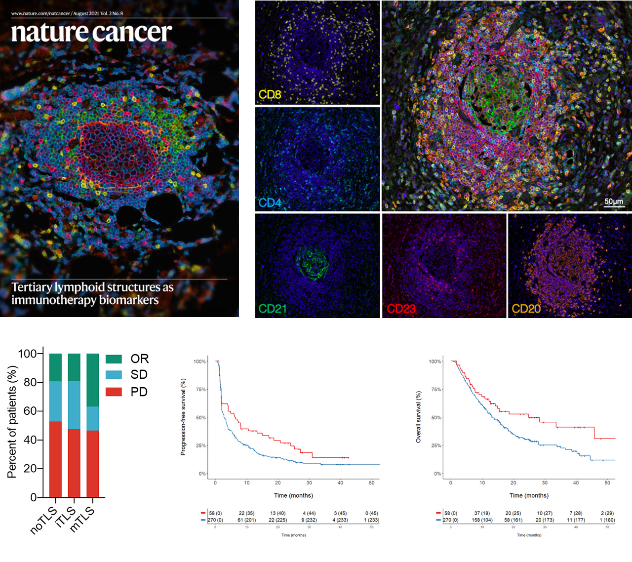
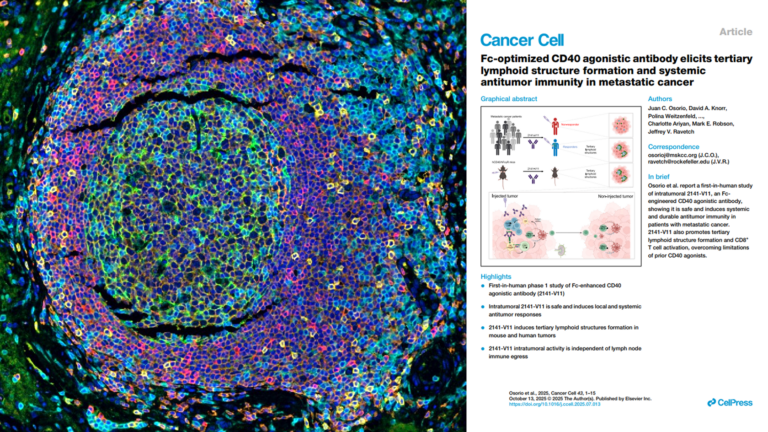
Mature TLS associated with improved ICI response and survival across solid tumors

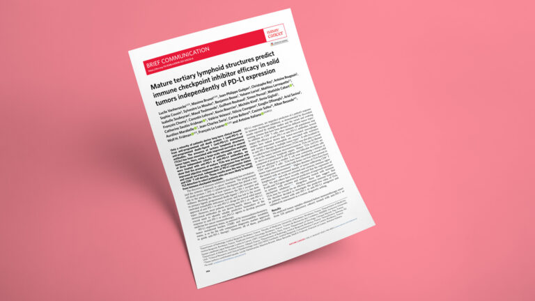
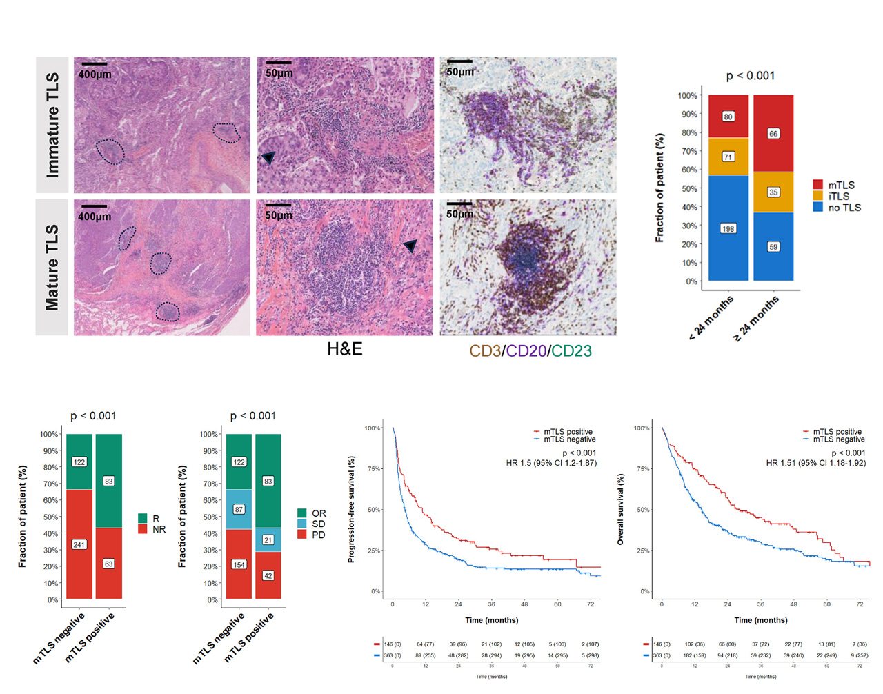
In three independent ICI-treated cohorts totaling 540 tumors (discovery n=328; validation n=131 and n=81), mature TLS were associated with improved objective response, progression-free survival and overall survival — independently of PD-L1 expression and CD8⁺ T-cell density.

Design: Retrospective pan-cancer analysis; three independent ICI-treated cohorts.

Endpoint: Objective response rate, progression-free survival, overall survival.

Independence: Associations held after adjustment for PD-L1 expression and CD8⁺ T-cell density.

Mature-TLS-predictive-biomarker-cancer-immunotherapy

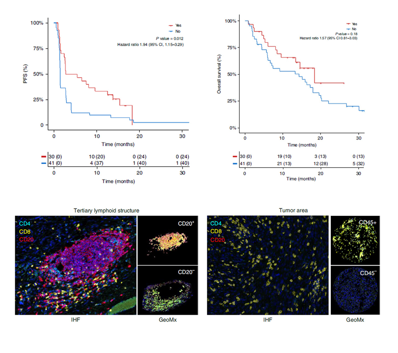
TLS-based selection was prospectively evaluated in a phase 2 sarcoma cohort

In the TLS-selected PEMBROSARC cohort (n=30), pembrolizumab plus low-dose cyclophosphamide achieved a 6-month non-progression rate of 40% and an objective response rate of 30% — higher than the previously reported all-comer cohorts from the same study (4.9% and 2.4%).

Design: Prospective phase 2 basket cohort; TLS-positive advanced soft-tissue sarcoma.

Selection: Enrichment on mature TLS status assessed by the multiplex IF workflow.

Outcome: Non-randomized comparison to previously reported all-comer sarcoma cohorts.

TLS-based selection was prospectively evaluated in a phase 2 sarcoma cohort

Mature TLS remained associated with ICI outcome in a 509-patient NSCLC cohort

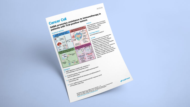
In 509 ICI-treated patients with NSCLC, mature TLS were associated with improved clinical outcomes — independently of PD-L1 expression and genomic features. Spatial transcriptomics and multiplex IF then identified tumor-microenvironment features associated with primary resistance within mTLS-positive tumors.

Cohort: 509 ICI-treated NSCLC patients.

Endpoint: Clinical outcome, independent of PD-L1 expression and genomic features.

Extension: Spatial transcriptomics + multiplex IF resolving primary-resistance features within mTLS-positive tumors.

Mature TLS remained associated with ICI outcome in a 509-patient NSCLC cohort

NEED A DIFFERENT TISSUE BIOMARKER ASSAY?

Develop and validate a custom IHC/IF assay for your trial

Explicyte develops and analytically validates custom IHC and multiplex IF tissue biomarker assays, from assay optimization through controlled clinical-trial deployment.

The Explicyte team at their Bordeaux laboratory

contact our team

Discuss TLS scoring for your clinical study

Tell us the indication, trial phase, tissue type, number of cases, available FFPE material, planned study timepoints and how TLS status will be used in the protocol. We’ll confirm sample requirements, reporting format, study workflow and timeline.

Answers about TLS scoring

Frequently asked questions

What samples can be analyzed with the TLS scoring assay?

The assay is designed for FFPE tumor tissue. Sponsors can provide FFPE blocks or unstained slides, with tissue and section requirements confirmed before study initiation. A pathology suitability review is performed before scoring to identify samples that may not provide sufficient evaluable tissue.

Each evaluable case receives a standardized TLS classification according to the validated scoring method, together with representative supporting images. Cohort-level summaries can also be provided when required by the study design.

TLS classification integrates tissue morphology, lymphoid organization and the CD4/CD8/CD20/CD21/CD23 multiplex IF profile. In the published workflow underlying the assay, mature TLS show germinal-center maturation, identified by a visible germinal center on HES and/or a CD23-positive follicular dendritic-cell network, whereas immature TLS lack these maturation features. The commercial assay is interpreted according to its controlled scoring rubric.

TLS status can be incorporated into a clinical-study biomarker strategy when supported by the protocol and development plan. Published studies have associated mature TLS with improved outcomes under immune-checkpoint blockade, and TLS-based enrichment was prospectively evaluated in the PEMBROSARC phase 2 cohort. The Explicyte assay is not, by itself, a CE-marked IVD or approved companion diagnostic; any formal patient-selection or regulated diagnostic strategy should be defined with the sponsor.

FFPE biopsies and resection specimens can be considered, provided sufficient evaluable tumor tissue is available. Because TLS are spatially heterogeneous structures, tissue amount and sampling context can affect their detection; sample adequacy and study design should therefore be reviewed before testing, particularly for small biopsies.

Turnaround depends on cohort size, sample format, tissue quality and reporting requirements. Share the number and type of samples with us and we will define the study schedule before initiation.

The current assay builds on the TLS classification strategy and CD4/CD8/CD20/CD21/CD23 multiplex IF approach used in Explicyte-supported publications. It is now implemented as a controlled assay under Explicyte’s ISO-certified quality-management framework.

Explicyte Oncology CRO logo

Capabilities

Modalities