Digital pathology CRO services: IHC, multiplex IF and quantitative histopathology

Explicyte operates an integrated digital pathology platform in Bordeaux, France, supporting academic, biotech and pharmaceutical teams from tissue preparation and pathologist-guided assay design to automated IHC/multiplex IF staining, whole-slide imaging and quantitative analysis.

With 150+ tumor and immune markers and 120+ IHC/mIF panels, we support tissue-based biomarker studies from target characterization and immune profiling to translational research and clinical development. Pathologist-guided scoring and in-house quantitative image analysis turn stained slides into interpretable, decision-ready datasets.

Explicyte delivers end-to-end digital pathology studies from tissue preparation and IHC/multiplex IF staining to whole-slide imaging, biomarker scoring and quantitative spatial analysis, with pathology input available where relevant.

Our mIF / IHC data have been published in

Cancer Cell journal logoClinical cancer research journal logoScience Advances logoCancer Cell journal logoSignal transduction and targeted therapy journal logo
Cancer Cell journal logoClinical cancer research journal logoScience Advances logoCancer Cell journal logo
Cancer Cell journal logoClinical cancer research journal logoScience Advances logoCancer Cell journal logoSignal transduction and targeted therapy journal logoNature cancer journal logo

Digital pathology CRO services

Why teams run digital pathology with us

ISO-certified pathology workflows

ISO 13485:2016 and ISO 9001:2015 certified for tissue-biomarker development and testing, with dedicated workflows for clinical-stage pathology studies.

A pathology platform built for fast turnaround

Two automated staining systems and two multispectral imaging systems, combined with in-house image analysis, minimize bottlenecks from staining to quantitative results.

Established immuno-oncology expertise

150+ validated tumor and immune markers, 120+ IHC/mIF panels, and a peer-reviewed publication track record across translational oncology.

Explicyte is certified ISO 9001:2015 and ISO 13485:2016 by Euro-Quality System (certificates 260133/1637F/1 and 260133/1637F/2). Scope: Precision Oncology — design and development of tissue biomarkers, and tissue biomarker testing services to support therapeutic decision-making. The certification governs our histopathology workflow; the image-analysis pipeline is delivered by the same team but sits outside the certified scope.

Explore our ISO-certified pathology services

HOW WE RUN DIGITAL PATHOLOGY STUDIES

From tissue to quantitative biomarker data

  1. Digital pathology study design and tissue strategy
    1

    Objectives, cohort and biomarker strategy

    We start from the biological or clinical question, available samples and intended readouts. Together we define tissue eligibility, markers and controls, single-plex or multiplex format, scoring strategy and quantitative analysis plan, with pathologists inputs incorporated where relevant.

  2. FFPE tissue processing and IHC multiplex IF assay development
    2

    Tissue processing and assay setup

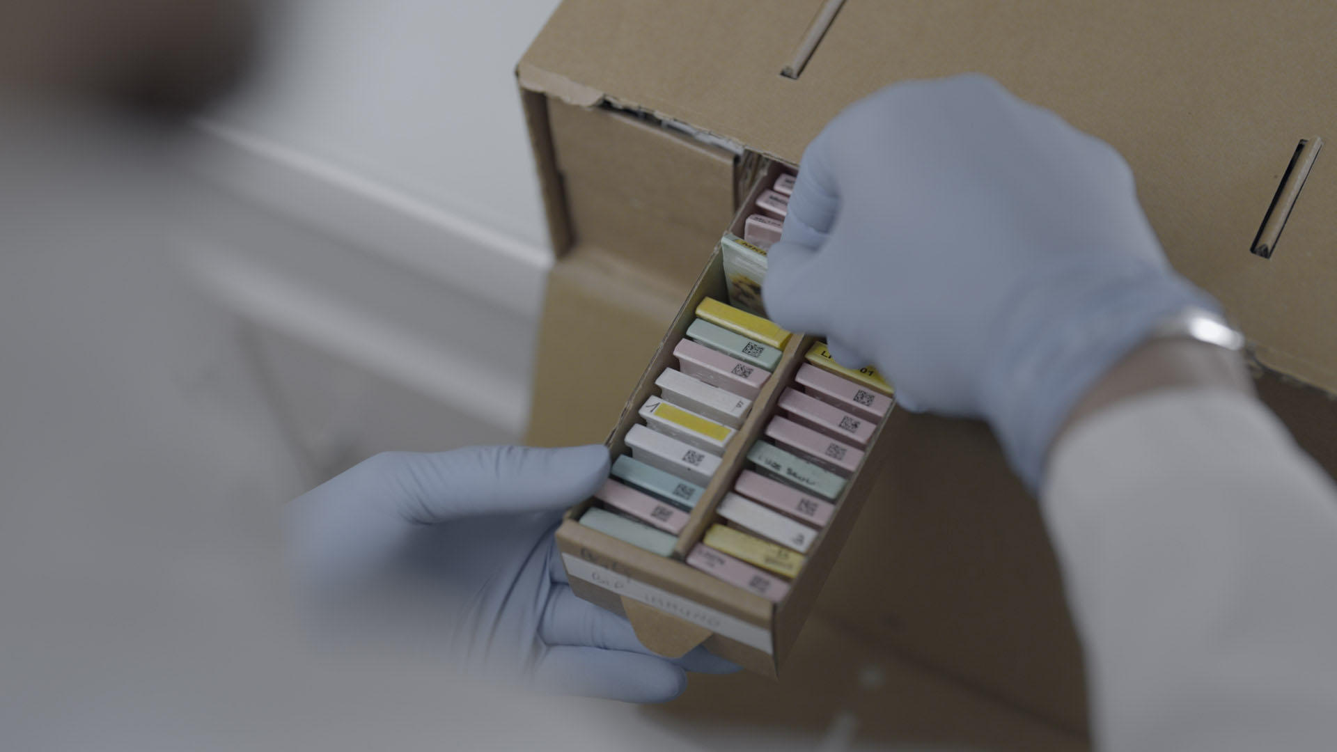
    We can work from sponsor-provided material or support human tissue sourcing where needed. In-house preparation can include fixation, embedding, sectioning, H&E and slide preparation. Marker conditions and controls are optimized before antibodies are incorporated into single-plex IHC or multiplex IF assays.

  3. Automated IHC multiplex IF staining and digital slide imaging
    3

    Automated staining and digital imaging

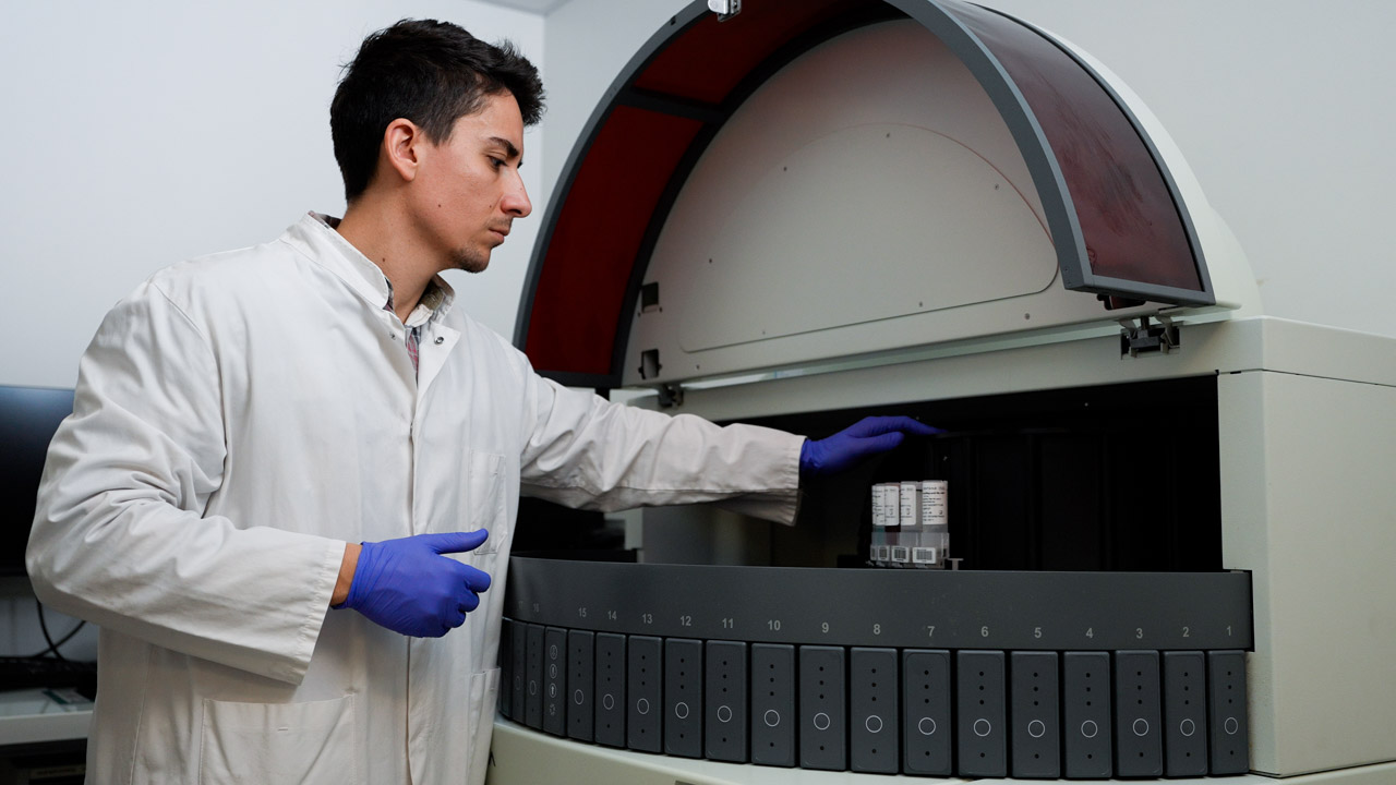
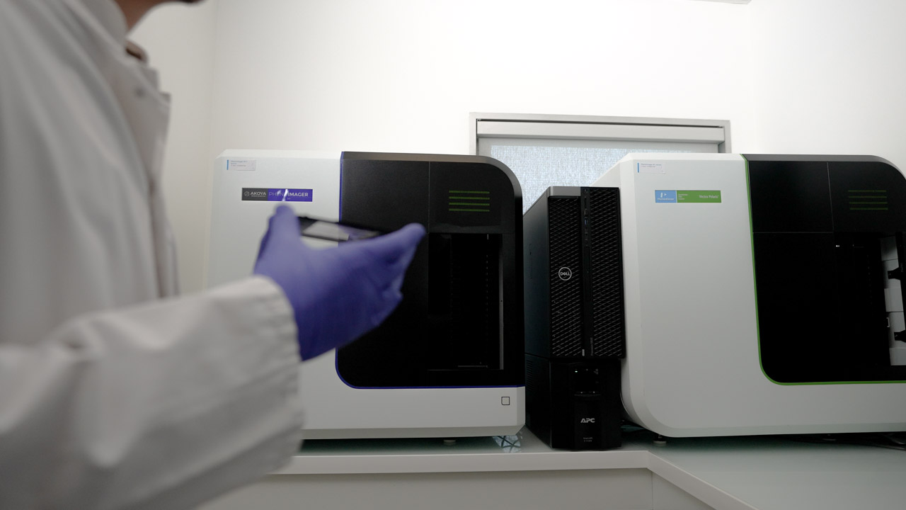
    IHC and multiplex IF assays are run on automated Ventana Discovery systems. Slides are digitized on Quanterix multispectral imaging systems, with image and staining QC performed before quantitative analysis or pathologist scoring.

  4. Quantitative digital pathology image analysis and interpretation
    4

    Quantification, scoring and interpretation

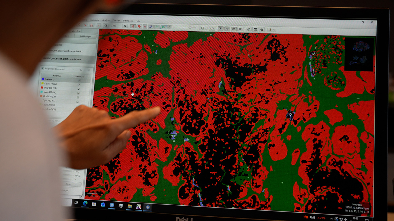
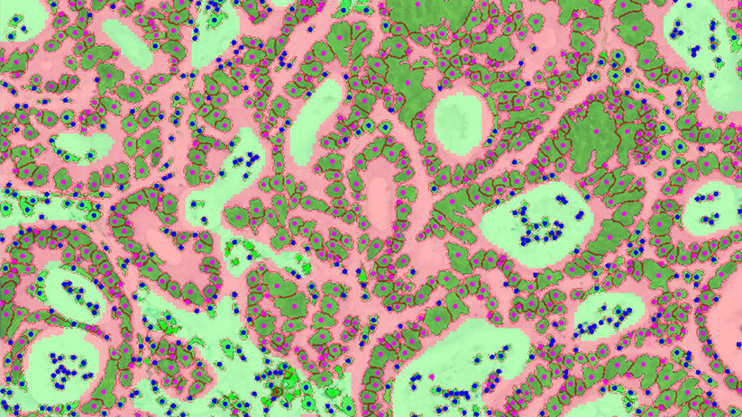
    Depending on study scope, readouts can include pathologist scoring and/or in-house quantitative image analysis, including tissue and cell segmentation, marker expression, cell phenotyping, compartment-specific densities and spatial relationships. Results are delivered as structured data, figures and study-specific reporting..

DIGITAL PATHOLOGY AT A GLANCE

IHC, multiplex IF & quantitative pathology capabilities

Sample types
FFPE and frozen tissue, biopsies, surgical specimens and TMAs; human tumor/normal tissue and samples from preclinical models.
Pathology
H&E review, tissue qualification, ROI annotation, biomarker scoring and study-specific pathology review, where required.
Staining
Automated single-plex IHC and multiplex immunofluorescence on two Ventana Discovery systems, with custom assay and panel development.
Marker library
150+ tumor and immune markers and 120+ IHC/mIF panels across translational oncology applications.
Digital imaging
Two Quanterix multispectral imaging systems for high-throughput digitization of multiplex pathology slides.
Quantitative analysis
Pathologist-guided and AI-assisted tissue/cell segmentation, phenotyping, marker quantification and spatial analysis through our in-house data science team.

What you receive

  • Digitized H&E, IHC and/or multiplex IF images according to study scope
  • Assay conditions, staining QC and panel documentation
  • Pathologist annotations and biomarker scores where included
  • Cell- and ROI-level quantitative tables for marker expression and cell phenotypes
  • Tissue-compartment maps and quantitative summaries
  • Spatial metrics and between-group comparisons where included in the analysis plan
  • A results session with your study director
  • Structured study outputs and publication-ready figures

DIGITAL PATHOLOGY CASE STUDIES

Digital pathology in practice
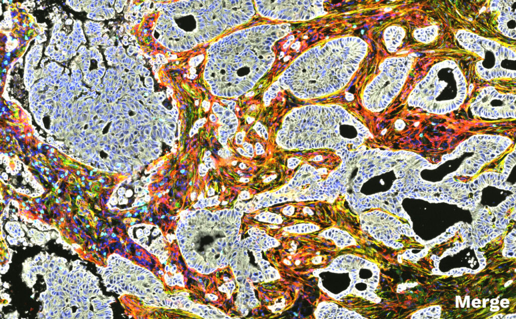
From cell phenotype to spatial relationships: multiplex digital pathology in NSCLC

Using multiplex immunofluorescence and quantitative image analysis, Explicyte characterized immune-exhaustion phenotypes, cancer-associated fibroblast populations and their spatial organization wit…

Multiplex digital pathology reveals immune exhaustion, stromal phenotypes and spatial organization in NSCLC

Histology services (IHC/IHF) : 150 validated markers, 120+ panels for Immuno-Oncology

Get our list of validated markers & panels (IHC/IHF) for immuno-oncology

Histology services (IHC/IHF) : 150 validated markers, 120+ panels for Immuno-Oncology

Quantitative histology to characterize immune contexture in human lung adenocarcinoma samples

The tumor microenvironment plays a central role in cancer progression and response to immunotherapy. Immune checkpoint inhibitors targeting the PD-1/PD-L1 axis and CTLA-4 have demonstrated major cl…

Quantitative histology to characterize immune contexture in human lung adenocarcinoma samples

B Cells and Tertiary Lymphoid Structures as Key Players in Anti-Tumor Immune Response

B cells are increasingly recognized as key contributors to anti-tumor immune responses and response to cancer immunotherapy. Recent studies have demonstrated the association between B cell-enriched…

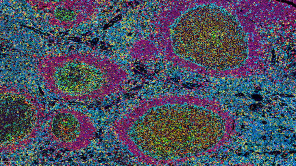
Case study: Multiparametric TLS characterization by digital pathology

DIGITAL PATHOLOGY PUBLICATIONS

Peer-reviewed studies using Explicyte IHC & multiplex IF data

GABA promotes resistance to immunotherapy in patients with TLS-positive tumors
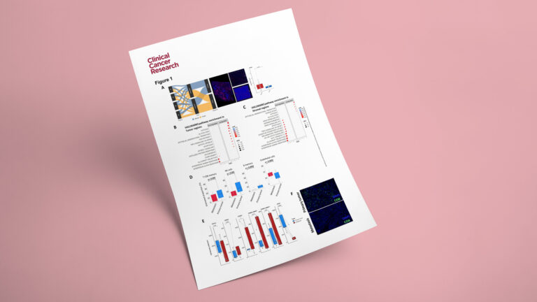
GABA promotes resistance to immunotherapy in patients with TLS-positive tumors

2026
Adenosine A2B Receptor Promotes Tumor Progression and Metastases in Undifferentiated Pleomorphic Sarcoma

Adenosine A2B Receptor Promotes Tumor Progression and Metastases in Undifferentiated Pleomorphic Sarcoma

2026
Immune control of functional memory CD8 T cells in normal-appearing vitiligo skin
Immune control of functional memory CD8 T cells in normal-appearing vitiligo skin

2025
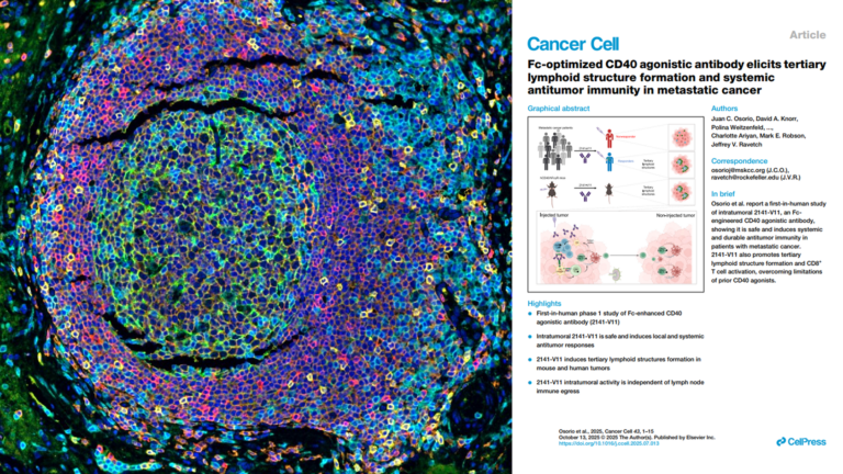
Fc-optimized CD40 agonistic antibody elicits tertiary lymphoid structure formation and systemic antitumor immunity in metastatic cancer

Fc-optimized CD40 agonistic antibody elicits tertiary lymphoid structure formation and systemic antitumor immunity in metastatic cancer

2025
Reshaping the tumor microenvironment of cold soft-tissue sarcomas with anti-angiogenics: a phase 2 trial of regorafenib combined with avelumab
Reshaping the tumor microenvironment of cold soft-tissue sarcomas with anti-angiogenics: a phase 2 trial of regorafenib combined with avelumab

2025
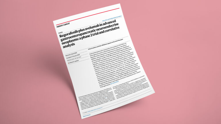
Regorafenib plus avelumab in advanced gastroenteropancreatic neuroendocrine neoplasms: a phase 2 trial and correlative analysis


Regorafenib plus avelumab in advanced gastroenteropancreatic neuroendocrine neoplasms: a phase 2 trial and correlative analysis

2025
Spatially resolved transcriptomics reveal the determinants of primary resistance to immunotherapy in NSCLC with mature tertiary lymphoid structures


Spatially resolved transcriptomics reveal the determinants of primary resistance to immunotherapy in NSCLC with mature tertiary lymphoid structures

2025
Tertiary lymphoid structures and cancer immunotherapy: From bench to bedside
Tertiary lymphoid structures and cancer immunotherapy: From bench to bedside

2025
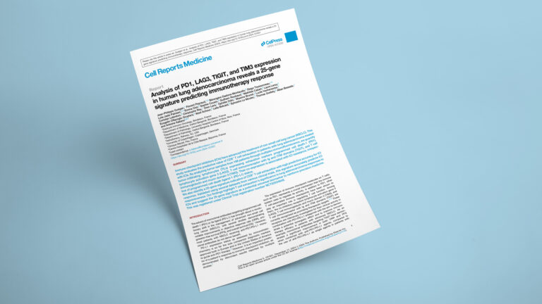
Analysis of PD1, LAG3, TIGIT, and TIM3 expression in human lung adenocarcinoma reveals a 25-gene signature predicting immunotherapy response


Analysis of PD1, LAG3, TIGIT, and TIM3 expression in human lung adenocarcinoma reveals a 25-gene signature predicting immunotherapy response

2024
Predictive value of tumor microenvironment on pathologic response to neoadjuvant chemotherapy in patients with undifferentiated pleomorphic sarcomas

Predictive value of tumor microenvironment on pathologic response to neoadjuvant chemotherapy in patients with undifferentiated pleomorphic sarcomas

2024
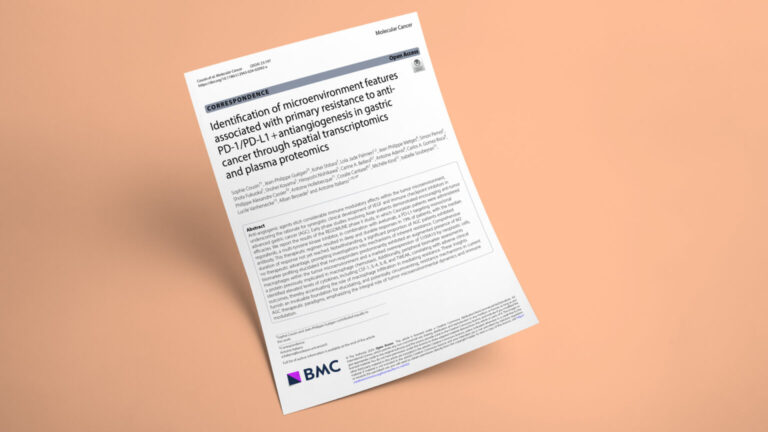
Identification of microenvironment features associated with primary resistance to anti-PD-1/PD-L1 + antiangiogenesis in gastric cancer through spatial transcriptomics and plasma proteomics

Identification of microenvironment features associated with primary resistance to anti-PD-1/PD-L1 + antiangiogenesis in gastric cancer through spatial transcriptomics and plasma proteomics

2024
Deciphering the correlation between metabolic activity through 18F-FDG-PET/CT and immune landscape in soft-tissue sarcomas: an insight from the NEOSARCOMICS study

Deciphering the correlation between metabolic activity through 18F-FDG-PET/CT and immune landscape in soft-tissue sarcomas: an insight from the NEOSARCOMICS study

2024
explicyte multiomics transcriptomics CRO team

Paul Marteau, PharmD (study director), Imane Nafia, PhD (CSO), Loïc Cerf, MSc (COO), Alban Bessede, PhD (founder, CEO), Jean-Philippe Guégan, PhD (CTO)

Contact our team

Discuss your digital pathology project

Tell us your tissue type, cohort, biomarkers and required readouts. We'll help define the staining, imaging, pathology and quantitative-analysis workflow for your study.

Answers about digital pathology services

Frequently asked questions

What digital pathology services does Explicyte provide?

Explicyte supports digital pathology studies from tissue sourcing and preparation through IHC/multiplex IF staining, digital imaging, biomarker scoring and quantitative image analysis. Projects can be run end to end or scoped to individual workflow steps.

We work with FFPE and frozen tissue, biopsies, surgical specimens and TMAs from human tumor or normal tissue and from preclinical models. We can also support custom sourcing of human tumor tissue through accredited French biobanks.

Yes. We optimize markers, controls and staining conditions before combining antibodies into study-specific panels. Our existing marker and panel library can provide a starting point for custom studies. For clinical-stage biomarker programs requiring controlled assay development and validation, we offer dedicated ISO-certified pathology services.

Pathology input can be incorporated where relevant, including sample qualification, H&E review, ROI annotation, biomarker scoring and review of specific tissue structures or compartments such as TLS.

Yes. Image analysis is available as a standalone service for digitized IHC and multiplex IF slides generated by Explicyte, another CRO or an internal laboratory. See our dedicated mIF & IHC image analysis services page for preprocessing, phenotyping and advanced spatial-analysis options.
Depending on the study design, outputs can include marker intensity and positivity, cell counts and densities, cell phenotypes, tumor-versus-stroma comparisons, biomarker scores, tissue-compartment maps and spatial relationships between cell populations. Advanced spatial analysis is scoped through our dedicated image-analysis workflow.
Yes. For clinical-stage programs requiring a defined quality framework, Explicyte provides dedicated ISO-certified pathology services covering IHC/IF assay development, assay validation and multiplex IF TLS scoring. These services are described separately in our Oncology Trials section.

Digital pathology measures tissue morphology and protein biomarkers using IHC or multiplex IF, making it particularly useful for target validation, cell phenotyping, biomarker scoring and spatial protein relationships. Spatial transcriptomics measures spatial RNA expression and is better suited to broad transcriptomic discovery or, depending on the platform, direct cell-level gene-expression localization. Many translational programs combine both approaches.

Explicyte Oncology CRO logo

Capabilities

Modalities