
Indoleamine 2,3-dioxygenase 1 is expressed in tertiary lymphoid structures and associated with improved outcome in patients with advanced non-small cell lung cancer treated with anti-PD1/PDL1 inhibitors
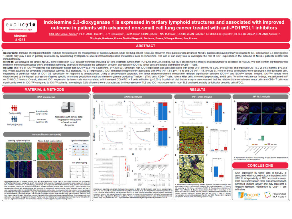
Background Immune checkpoint inhibitors have revolutionized the management of patients with non-small cell lung cancer. However, most patients with advanced NSCLC display primary resistance to these treatments. Indoleamine 2,3-dioxygenase 1, or IDO1, may contribute to immune regulation by catabolizing tryptophan into several immunosuppressive metabolites, including kynurenine. This study aimed to investigate the role of IDO1 […]
How to screen for mature tertiary lymphoid structures in routine pathology — a standardized method validated across 357 tumors
Why intravenous oncolytic virus JX-594 lit up immune biomarkers but not tumor responses in HR+ breast cancer

Why the TLR4 agonist G100 boosted T cells but not PD-1 response in “cold” soft tissue sarcoma

Why systemic oncolytic virus JX-594 stirred an immune signal but not clinical benefit in soft tissue sarcoma

Can a simple blood test predict who responds to immunotherapy? Circulating L-arginine tracks checkpoint-inhibitor survival

Identification of super-exhausted T cells: a novel population predictive of response to immunotherapy
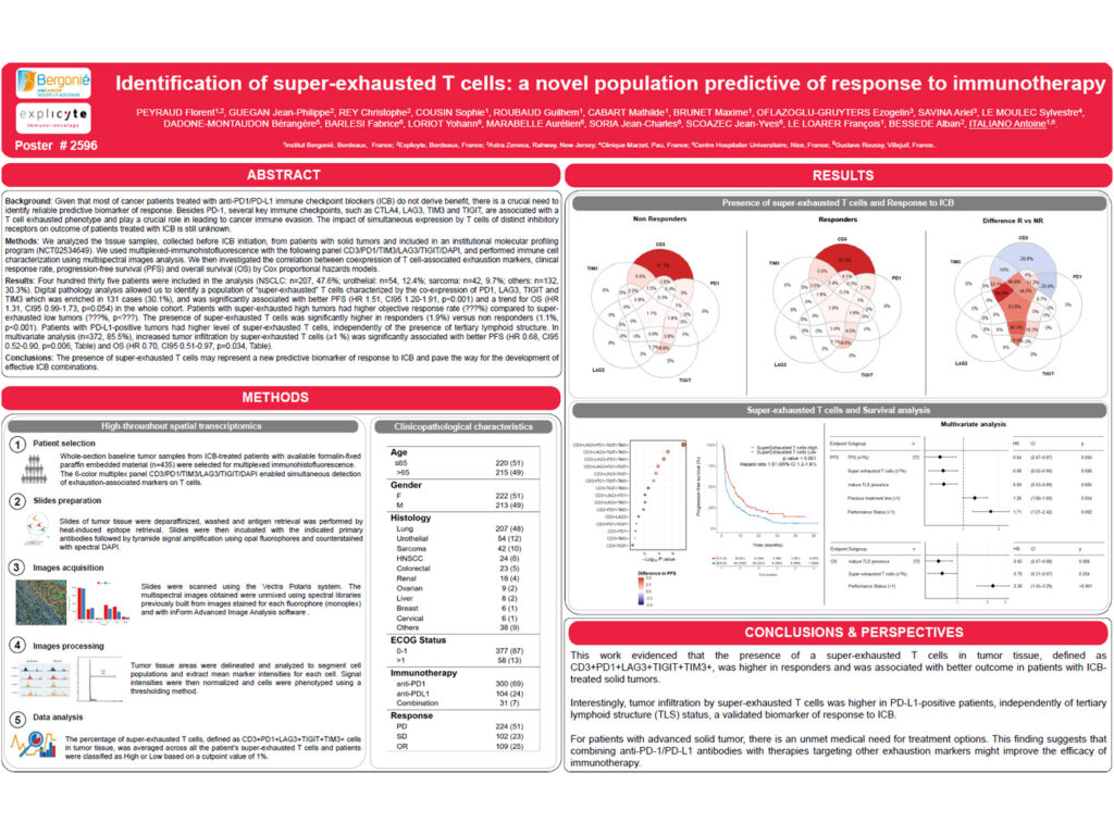
Background Most cancer patients treated with anti-PD1 or anti-PDL1 immune checkpoint blockers do not derive clinical benefit. There is therefore a crucial need to identify reliable predictive biomarkers of response. Beyond PD1, several immune checkpoints, including CTLA4, LAG3, TIM3 and TIGIT, are associated with a T cell exhausted phenotype and play an important role in […]
Does acetaminophen blunt cancer immunotherapy? Plasma evidence links paracetamol to worse checkpoint-inhibitor outcomes

How tertiary lymphoid structures predict pembrolizumab response in soft-tissue sarcoma — and why plasma cells matter
Ovarian cancer ascites display altered immune environment featured by enhanced soluble factors and suppressive cellular context
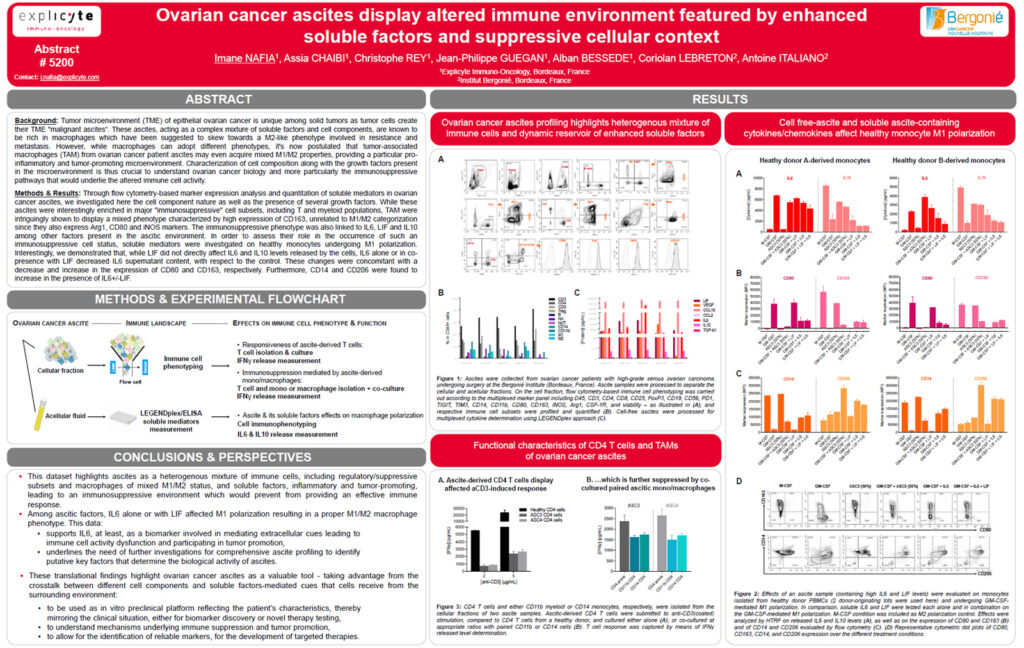
Background The tumor microenvironment of epithelial ovarian cancer is unique among solid tumors, as tumor cells can shape their own microenvironment through the formation of malignant ascites. These ascites represent a complex mixture of soluble factors and cellular components. They are known to be enriched in macrophages, which have been suggested to acquire an M2-like […]